Introduction
Spinal stenosis is a condition that occurs when the spinal canal narrows, leading to compression of the spinal cord and nerves. This narrowing can happen anywhere in the spine, though it is most commonly seen in the neck (cervical stenosis) and lower back (lumbar stenosis). Spinal stenosis may cause a variety of symptoms such as pain, numbness, weakness, and tingling, affecting the mobility and daily functioning of the individual. Early diagnosis and intervention are crucial in managing the condition effectively.
1. Medical History and Symptom Review
The first step in spinal stenosis diagnosis is a thorough review of the patient’s medical history and symptoms. Understanding the patient’s symptoms, medical background, and any risk factors helps guide further diagnostic steps.
a. Symptom Overview:
Spinal stenosis can present with a range of symptoms, which may vary depending on the location of the narrowing. Common symptoms include:
-
Pain :- Patients may experience localized or radiating pain in the back, neck, or limbs. The pain can be aggravated by activities such as walking or standing for prolonged periods.
-
Numbness and Tingling :- A feeling of numbness or a “pins and needles” sensation may be felt in the affected limbs, particularly in the legs or arms.
-
Weakness :- Muscle weakness can develop due to nerve compression, making it difficult to walk or perform daily tasks.
-
Bladder or Bowel Dysfunction :- In severe cases, spinal stenosis in the lumbar region may cause issues with bowel and bladder control due to pressure on the nerves controlling these functions.
-
Postural Changes :- Some patients may notice that bending forward or sitting down relieves their symptoms, while standing or walking may worsen them.
A detailed discussion of these symptoms with the healthcare provider is essential in determining whether spinal stenosis could be the cause. The physician will inquire about the onset, intensity, and duration of the symptoms, as well as any contributing factors such as physical activity or injury.
b. Risk Factors to Consider:
-
Age :- Spinal stenosis is more common in older adults due to the natural wear and tear of the spine.
-
Genetic Factors :- In some cases, spinal stenosis is congenital (present at birth) and may be due to abnormal spinal development.
-
Previous Injuries :- Past spinal injuries, such as fractures or herniated discs, can contribute to the development of spinal stenosis.
-
Osteoarthritis :- Degenerative changes in the spine, such as bone spurs and thickened ligaments, often contribute to the narrowing of the spinal canal.
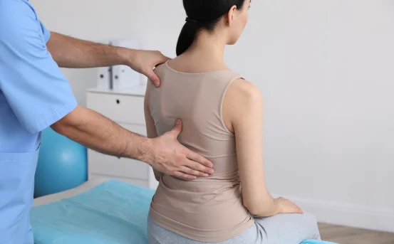
2. Physical Examination
Once the patient’s history has been reviewed, a physical examination is conducted to evaluate the patient’s condition. The physical exam focuses on assessing the patient’s neurological function and identifying any signs of nerve compression.
a. Neurological Assessment:
During the examination, the physician will assess various aspects of neurological function, including:
-
Muscle Strength :- The physician will test the strength of different muscle groups in the arms and legs to detect any signs of weakness.
-
Sensation :- Sensory testing is conducted to check for areas of numbness or abnormal sensations, such as tingling or a “pins and needles” feeling.
-
Reflexes :- The physician will check for abnormal reflexes, such as diminished or absent reflexes, which may indicate nerve compression.
b. Spinal Alignment and Mobility
The doctor will also check the alignment of the spine and assess the patient’s range of motion. Limited mobility, pain, or tenderness during movement may indicate the presence of spinal stenosis.
-
Straight Leg Raise Test :- This test helps assess nerve root involvement in the lower back. When a patient lies on their back and the leg is raised, the physician observes whether the patient experiences pain or discomfort, which could indicate nerve compression.
-
Posture Evaluation :- Posture-related changes, such as a forward-leaning posture or the inability to stand upright for long periods, may also suggest spinal stenosis.
3. Imaging Tests
If spinal stenosis is suspected based on the patient’s medical history and physical examination, imaging tests are typically used to confirm the diagnosis and evaluate the severity of the condition. These tests provide detailed images of the spine and help doctors determine the exact location of the narrowing.
a. X-rays :- X-rays are often the first imaging test used to evaluate the spine. While X-rays do not provide detailed images of the spinal cord or nerves, they can help identify bone spurs, degenerative changes, or misalignments in the vertebrae. X-rays can also rule out other conditions such as fractures or tumors that may mimic the symptoms of spinal stenosis.
X-rays can show:
- Disc space narrowing
- Bone spurs (osteophytes)
- Spondylolisthesis (vertebral slippage)
However, X-rays are limited in their ability to detect soft tissue abnormalities such as nerve compression.
b. Magnetic Resonance Imaging (MRI)
MRI is the gold standard for diagnosing spinal stenosis. It provides detailed images of the spinal cord, nerve roots, and soft tissues, allowing doctors to identify areas of nerve compression, bulging discs, and other spinal abnormalities.
MRI scans are non-invasive and offer high-resolution images, making them especially useful for evaluating the severity of stenosis and the degree of pressure on the spinal cord and nerves.
MRI can detect:
- Bulging or herniated discs
- Thickened ligaments
- Tumors or cysts (although rare in spinal stenosis)
- Spinal cord or nerve root compression
c. CT Myelography
CT myelography is a more advanced imaging test that combines a CT scan with an injection of contrast dye into the spinal fluid. This test allows for a detailed view of the spinal cord, nerve roots, and surrounding structures. CT myelography is typically used if an MRI is unavailable or inconclusive.
4. Electromyography (EMG) and Nerve Conduction Studies
In some cases, electromyography (EMG) and nerve conduction studies may be conducted to assess the function of the nerves and muscles. These tests help doctors evaluate whether nerve compression is causing weakness or abnormal sensations in the limbs.
-
EMG measures electrical activity in muscles, which can help identify areas of nerve damage.
-
Nerve conduction studies assess how well electrical signals travel along the nerves, helping identify nerve damage caused by spinal stenosis.
These tests are particularly helpful when there is a concern about muscle weakness or sensory disturbances that could be attributed to nerve compression.
5. Differential Diagnosis
In some cases, other conditions may mimic the symptoms of spinal stenosis. Therefore, it is important for doctors to rule out other possible causes of the patient’s symptoms. Conditions that may be considered in the differential diagnosis include:
-
Herniated Disc :- A herniated disc can also cause nerve compression and similar symptoms to spinal stenosis.
-
Spondylolisthesis :- This condition involves the slippage of one vertebra over another and can result in symptoms similar to those of spinal stenosis.
-
Arthritis :- Osteoarthritis can cause narrowing of the spinal canal and lead to similar symptoms, especially in older adults.
-
Tumors or Infections :- Although rare, tumors or infections in the spinal cord can also cause narrowing of the spinal canal and nerve compression.
Conclusion
The diagnosis of spinal stenosis involves a thorough review of the patient’s medical history, a detailed physical examination, and advanced imaging studies. MRI is the primary diagnostic tool for visualizing soft tissue changes and nerve compression, while X-rays provide a view of bone structures. Electromyography and nerve conduction studies may be used in cases where nerve damage is suspected.
If you experience symptoms such as back pain, leg weakness, numbness, or difficulty walking, it is essential to seek medical attention promptly. Early diagnosis and treatment can significantly improve outcomes and help manage the symptoms of spinal stenosis effectively, preventing further complications and improving your quality of life.