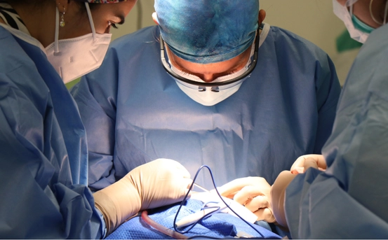
The retina is a sophisticated, light-sensitive tissue lining the back of the eye—essentially our biological camera sensor. When this retina becomes damaged—due to tears, holes, detachments, traumatic injuries, growths, or disease—vision can deteriorate quickly, sometimes irreversibly. This is where retinal surgery plays a life-changing role.
In modern ophthalmology, several advanced techniques exist to address a wide spectrum of retinal conditions. From minimally invasive laser procedures to intricate microsurgeries, each method has specific indications, benefits, and considerations. This comprehensive guide explores the most common types of retinal surgery—pneumatic retinopexy, scleral buckle, vitrectomy, laser photocoagulation, and select specialized procedures—along with when and why they’re used. Understanding these types of retinal surgery can help patients make informed decisions about their treatment options.
Types of Retinal Surgery
In this article, we will delve into the various types of retinal surgery available, emphasizing their purposes and techniques.
Pneumatic Retinopexy
- What it is: An in‑office procedure where a gas bubble (like SF₆ or C₃F₈) is injected into the eye. The bubble presses the retina against its natural position while laser or freeze treatments seal off accompanying retinal tears.
- Ideal for: Small, uncomplicated, superior retinal detachments with a single or limited tear.
Pros & Cons
- Minimally invasive, typically outpatient
- Requires strict head positioning post‑op, and no flying or altitude changes until the bubble resorbs.
Scleral Buckle
- What it is: A silicone band or sponge is sutured around the eyeball, indenting its wall, allowing the retina to reattach, often combined with cryotherapy or laser sealing.
- Ideal for: Chronic detachments, multiple breaks, inferior detachments, or in younger, phakic (naturally lens‑intact) patients.
Pros & Cons
- Permanent support to the retinal break
- Minor surgery with potential infection, inflammation, or postoperative diplopia.
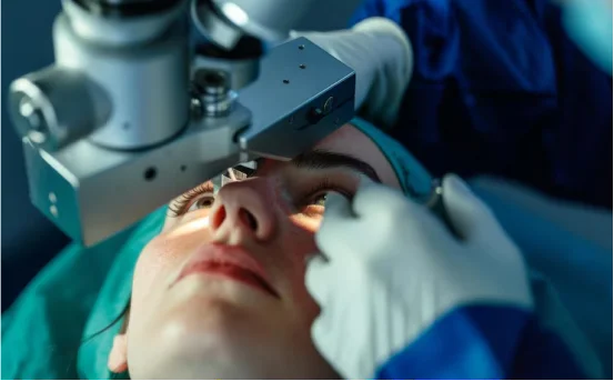
Vitrectomy
- What it is: Microsurgical removal of the vitreous gel through micro-incisions (23–27 gauge). The surgeon can then drain subretinal fluid, relieve traction, and seal tears. A gas bubble or silicone oil may be inserted to support the retina.
- Ideal for: Complex or giant retinal tears, proliferative vitreoretinopathy, diabetic retinopathy complications, vitreous hemorrhage, macular hole, and more.
Pros & Cons
- Highly effective—primary reattachment success rates > 90%
- Can accelerate cataract formation, involve longer recovery, and carry added surgical risks .
Laser Photocoagulation (Laser Coagulation)
- What it is: Focused laser “burns” create adhesion around retinal tears or seal leaky vessels in diabetic or vascular eye disease.
- Ideal for: Retinal tears, diabetic retinopathy, macular edema, and some vein occlusions .
Pros & Cons
- Office-based, quick, and effective for focal issues
- Potential side effects include mild vision loss, blurred vision, or decreased night vision.
Anterior Vitrectomy
- What it is: Removal of vitreous gel from the front (anterior) portion of the eye, typically used when vitreous blocks the pupil or leaks out during cataract or glaucoma procedures.
- Ideal for: Especially useful when vitreous prolapse occurs during cataract removal, lens trauma, or glaucoma surgery.
Pros & Cons
- Prevents vitreous traction complications
- Requires precision due to delicate structures up front.
Specialized Procedures
- Scleral Reinforcement Surgery: Supportive surgery for high myopia, reducing progression of posterior staphyloma by reinforcing the sclera.
- Retinal Prosthesis and Translocations (rare, experimental): Include technologies like Argus II (retinal implants) and macular translocation, used in end-stage blindness or macular degeneration . These are generally not mainstream yet, but under study and early adoption.
Why Retinal Surgery Matters
Preservation and Restoration of Vision :- Untreated retinal tears or detachment can lead to permanent vision loss. Procedures like vitrectomy, pneumatic retinopexy, and scleral buckle can save at-risk vision by re‑attaching the retina and restoring blood flow.
Tailored Treatment for Diverse Conditions :- Whether it’s a single tear, diabetic hemorrhage, macular hole, or traumatic injury, modern retinal surgery offers precise solutions. Advances in micro‑incision instruments, enhanced visualization, and intraoperative OCT make it possible to treat a wide array of issues with accuracy and reduced collateral damage.
Rapid Technological Progress :- Microincisional vitrectomy systems (23–27 gauge), 3D visualization platforms, illuminated laser probes, and multifunctional instruments have transformed outcomes, patient comfort, and recovery times. As a result, success rates have soared and complexities previously deemed high risk are now routinely manageable.
Timely Intervention is Key :- Retinal detachments are vision emergencies. Delay by even 24 hours increases risk of permanent damage, especially to the macula. Awareness and prompt care are essential.
Who Should Consider Retinal Surgery?
- Individuals experiencing sudden flashes, floaters, shadow or curtain-like vision loss.
- Patients diagnosed with retinal tears, detachments, diabetic hemorrhages, macular holes, or epiretinal membranes.
- People who’ve already undergone cataract, glaucoma, or vitreoretinal procedures and experienced complications involving the vitreous or lens.
- Patients with progressive high myopia being evaluated for scleral reinforcement surgery.
Recovery and Post‑Op Considerations
- Positioning: Pneumatic retinopexy and vitrectomy (with gas/silicone) often require specific head positioning and avoidance of air travel until gas resorbs.
- Follow‑up: Regular follow-ups are essential to monitor retinal reattachment, detect cataract formation (common after vitrectomy), and ensure healing.
- Risks: Though success rates exceed 80‑90%, patients may still experience infection, hemorrhage, cataracts, increased intraocular pressure, double vision, or re-detachment—these are addressed promptly if they occur .
Conclusion
Retinal surgery today is a triumph of precision medicine. Techniques from laser sealing of tears to micro-incision vitrectomy and eye-wall buckling offer reliable, effective treatment for potentially sight‑threatening conditions. With technological innovation, surgeons can tailor interventions both to complexity and patient lifestyle—whether office-based pneumatic retinopexy or hospital-based vitrectomy.
At its core: retinal surgery is about preserving sight and improving quality of life. Quick action, informed patient choice, and cutting-edge surgical care combine to prevent vision loss, restore clarity, and help patients rediscover the world around them.
If you experience sudden visual disturbances—floaters, flashes, reduced vision—seek retinal specialist evaluation immediately. With timely care, most patients regain significant vision and confidence.