Retinal surgery is the set of operations conducted directly on the retina, which is the fragile light-sensitive tissue lining the inside rear of the eye. By turning incoming light into electrical impulses, the retina sends those signals along the optic nerve up to the brain for processing. Since any tear, detachment, or disease affecting this layer can quickly rob a person of clear sight, surgeons often move in to fix the damage. In simple terms, these procedures address specific problems, including retinal detachment, macular holes, diabetic retinopathy, and various vitreoretinal disorders. Because the field demands intense training and advanced tools, a successfully performed surgery can halt vision loss and may even restore sight that seemed permanently gone.
Retinal surgery is crucial for those experiencing severe vision problems.
Why Retinal Surgery Is Needed
Understanding the importance of retinal surgery can save a patient’s vision.
Retinal surgery steps in when a serious problem threatens sight unless specialists act quickly, so people keep as much usable vision as possible. Take a detached retina: because the delicate tissue will not fix itself, every minute counts, and vision can vanish overnight if the eye is ignored. Surgeons stress that a torn or lifted retina needs treatment as soon as possible, for waiting can lead to permanent blindness .The same sense of urgency surrounds advanced diabetic retinopathy, in which diabetes weakens retinal blood vessels, and macular holes-tears in the central retina-because medicated drops or even laser spots rarely solve the problem. Surgery then becomes crucial whenever retinal disease outpaces non-invasive care. Among the usual reasons for heading to the operating table are severe detachment, proliferative diabetic retinopathy, macular holes, epiretinal membranes, and other complicated conditions. Fortunately, todays procedures such as vitrectomy and scleral buckle achieve high success rates, reaching roughly ninety percent for rhegmatogenous detachments, yet timing remains the single most important factor in saving sight.
Patients experiencing symptoms need to consider retinal surgery without delay.
Symptoms That Suggest You Might Need Retinal Surgery
Patients often notice sudden changes in their vision that point to a serious problem in the retina. Classic signs of retinal detachment include new floaters-dark specks or threads that drift across sight-flashes of light, and a shadow or curtain slowly spreading over the field of view. The National Eye Institute, for instance, warns that a sudden rise in floaters, flashes of light, or a curtain or shadow over your vision represents these hallmark symptoms. The Cleveland Clinic echoes this, urging prompt medical attention for any new floaters, flashes, or darkening in the visual field. Other signs depend on the specific condition: a macular hole in the central retina can blur or warp straight-ahead vision-lines may bend-and eventually create a central blind spot, making reading hard. In diabetic retinopathy early stages are often symptom-free, yet later bleeding into the vitreous produces dark floaters or hazy sight. Any abrupt vision change-a gray curtain, blurry patch, or distortion-calls for an urgent dilated exam, because it could mean a tear or detachment that requires surgery.
Recognizing symptoms early can lead to timely retinal surgery interventions.
Causes of Retinal Disorders
Retinal surgery may be needed when damage beneath the eye’s surface threatens vision. Most often, the problem starts with a tear or a detachment. In rhegmatogenous detachment, age-related changes shrink the vitreous gel, which then pulls sharply on the retina. A tear opens, allowing fluid to slip below and lift the retina from its bed. Tractional detachment arises when scar tissue-hazardous growth from proliferative diabetic retinopathy-tugs the retina loose. The rarer exudative type develops when inflammatory fluid or leaky blood vessels collect under the retina, sometimes without any tear.
In many cases, retinal surgery is the only solution to restore vision.
Age and severe myopia-extreme nearsightedness-raise the odds of retinal detachment. The National Institutes of Health adds that significant eye trauma, prior procedures such as cataract extraction, and a relatives history of the problem increase vulnerability. Other risks include diabetic retinopathy, lattice degeneration (areas of weak, thin retina), posterior vitreous detachment, and various hereditary retinal disorders. For instance, diabetic retinopathy arises when chronically high blood sugar damages the vessels, allowing them to tear or pulling on the retina and causing a tractional detachment. In short, any condition that mechanically stretches, tears, or otherwise compromises the retinal surface-trauma, severe diabetes, or proliferative disease-can create the need for urgent surgical repair.
Conditions like diabetic retinopathy often result in the need for retinal surgery.
Diagnosis of Retinal Conditions
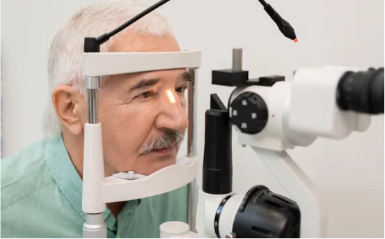
Diagnosing retinal problems begins with a thorough eye exam and specialized imaging. First the doctor performs a comprehensive, dilated eye exam in which drops widen the pupil so the full retina can be viewed with a slit lamp or ophthalmoscope. While the lids remain closed, the examiner may gently press on the eye to uncover hidden tears or early signs of detachment. If bleeding or opacities block the view, a handheld ultrasound may be substituted to scan the rear of the eye. Optical Coherence Tomography (OCT) is often added; this fast, noninvasive scan slices the retina into cross-sectional pictures. The detailed images reveal subtle defects such as small tears, detachment pockets, macular holes, or diabetic swelling that might be missed with a mirror alone. For example, OCT not only confirms a detachment or hole, but also outlines its size and location, making it key for tracking changes in diabetic retinopathy. If circulation issues are suspected, fluorescein angiography may be performed, injecting a harmless dye into the bloodstream to highlight retinal vessels. Above all, regular dilated exams matter; catching even a tiny tear early gives the best chance to seal it before a full detachment occurs.
Comprehensive exams can identify issues that may require retinal surgery.
Treatment Options (Including Surgery)
Doctors can manage retinal disorders through a range of methods, from simple in-office fixes to more involved surgical procedures.
Timely treatment options may include retinal surgery when conditions worsen.
- Laser photocoagulation or cryopexy :- When a tear is small or a detachment just beginning, the physician may close the gap without major surgery. In photocoagulation, a laser seals the tear by creating tiny burns that form a scar and bond the retina to the eye wall. Cryopexy achieves the same goal with a freezing probe that welds the edges of the break together. Both techniques are usually performed in the clinic with only local anesthesia.
- Pneumatic retinopexy :- This is a gentle approach for select detachments. The surgeon injects a small gas bubble into the vitreous, and the patient tilts their head so the bubble presses against the lifted retina and flattens it. Laser or cryo treatment is then applied around the tear to secure the repair. Over several days the bubble disappears on its own. Although there are no sutures, successful recovery depends on following strict head positions. Patients must understand that retinal surgery has specific protocols for recovery.
- Scleral-buckle surgery :- The ophthalmic team gently places a soft silicone band around the white outer layer of the eye, the sclera. This band lightly indents the eyeball, easing tension on the retina and pressing the detached tissue back against its normal position. While in the same setting, the surgeon usually seals any identified tears by applying either a laser or a freezing probe. Although the eye itself continues healing, the buckle stays in place for life, quietly supporting the repaired retina.
- Vitrectomy :- Vitrectomy is the go-to procedure for major retinal detachments or tricky situations such as macular holes and severe vitreous bleeds. The surgeon makes a tiny incision, removes the cloudy vitreous gel, and gains clear sight of the retina. With that access, she can seal tears or peel away scarred membranes with either laser light or fine-forcep technique. Finally, the original gel is replaced by sterile salt water, oil, or gas, pressing the retina back onto the eye wall. The gas bubble dissolves on its own, while silicone oil must be scheduled for a second sweep if it refuses to budge.
Post-operative care is vital for the success of retinal surgery.
No surgery, however skillful, guarantees success without careful aftercare. Right after the operation, most patients wear a snug patch and begin a course of antibiotic and steroid drops to ward off infection and soothe swelling. Patients who received a gas bubble then follow strict head positioning, often face-down or tilted to one side, for several days so the bubble cradles the retina with steady pressure. Air travel, even at modest altitude, is banned during this phase because expanding gas could turn the sealed eye into a painful emergency. In the first few days mild discomfort, redness, tears, and blurred vision are normal; these symptoms fade as the eye gels again and clarity steadily returns over the weeks that follow.
After surgery, doctors tell patients to skip heavy lifting and hard physical work until the surgeon says its safe. Patients usually notice some vision improvement within four to six weeks, although the final stage of healing may take several months. Scheduled follow-up visits, during which the doctor examines the eye with dilating drops, are crucial for tracking recovery and spotting problems early.
Adhering to follow-up visits is crucial after retinal surgery.
Conclusion
Retinal surgery can protect sight that is threatened by serious disorders. Because the retina cannot repair itself after it is torn or detached, quick diagnosis and treatment matter. Eye specialists emphasize that new floaters, flashes of light, or dark shadows should prompt an immediate office visit. When surgery is performed soon, the retina can often be reattached successfully-approximately 90 percent of patients retain useful vision according to current studies. Likewise, modern techniques for diabetic retinopathy and other retinal diseases-laser therapy, intravitreal injections, and vitrectomy-can slow damage if begun early. Regular eye examinations for people at higher risk, combined with fast action when symptoms arise, represent the best way to maintain clear vision and favorable long-term results.
Ultimately, retinal surgery can make a significant difference in recovery.