


Vitrectomy surgery is an advanced eye operation in which the surgeon removes the vitreous humor, the clear, jelly-like gel that sits between the lens and the retina. Because the procedure requires specialized instruments and training, it is usually carried out in a hospital or an outpatient surgical center equipped for high-resolution retinal work. Surgeons turn to vitrectomy when serious problems in the vitreous or retina threaten vision and other treatments such as laser therapy or medication have proven inadequate.
Patients often wonder how a doctor decides that this delicate procedure is needed at all. The call for vitrectomy grows only after a thorough workup that may include a detailed slit-lamp exam, wide-field imaging, optical coherence tomography, and the practiced eyes of a retina specialist weighing each finding. Getting to that decision early can save precious vision and give the surgeon the best chance to restore it.
Why is Vitrectomy Surgery Performed?
Vitrectomy is rarely offered as a first-line fix; it is typically reserved for sight-threatening disorders that will worsen without surgical access. Before the blades touch the eye, the care team pursues a full history, reviews every scan, and confirms that drug therapies or office procedures have failed. Only then is the procedure scheduled, marking a turn from watchful waiting to the precise, controlled repair the eye urgently needs.
This detailed process aids in determining the necessity of vitrectomy surgery.
Common Conditions That May Require Vitrectomy:
- Retinal Detachment :- When the retina lifts away from the layer beneath it, vision may vanish almost at once. Surgery replaces the detached tissue and restores its proper position.
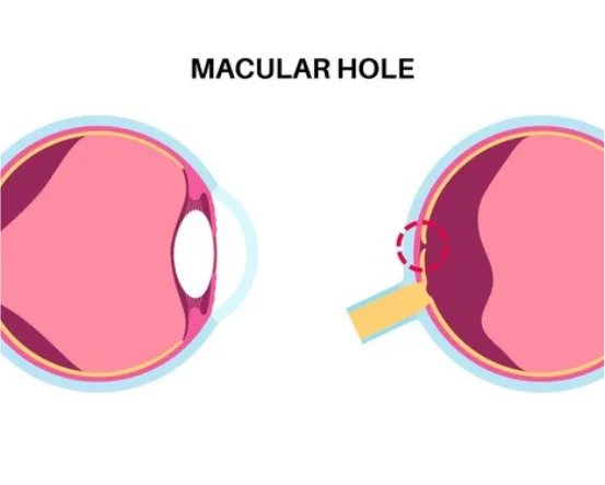
- Macular Hole :- Even a tiny rip in the macula can fog central sight. Removing cloudy vitreous and sealing the tear often sharpens or preserves vision.
- Diabetic Retinopathy :- In advanced stages, fragile vessels leak blood into the clear gel, blocking sight. Vitrectomy clears the gel, washes out the blood, and trims excess scar tissue.
- Eye Floaters & Vitreous Hemorrhage :- Stubborn floaters or bleeding shadows can crowd the field of vision. The procedure removes clouding gel and restores a clearer window.
- Infections (Endophthalmitis) :- Massive internal infections may spread quickly and prompt emergency surgery to evacuate infected fluid.
- Trauma or Injury to the Eye :- Serious blows, glass shards, or sports injuries may force surgeons to scoop out debris, leaking blood, and torn membranes.
Diagnosis for Vitrectomy: How is the Need Identified?
Consultation with a retina specialist is vital to assess the potential need for vitrectomy surgery.
Deciding on vitrectomy starts with a thorough history, careful exam, and images such as optical coherence tomography. Seeing problems early is vital to averting permanent vision loss.
A comprehensive evaluation often leads to the decision for vitrectomy surgery.
- Patient History and Symptom Assessment
Diagnosis starts with a careful discussion of symptoms and medical background. Warning signs that prompt immediate attention include:
- Sudden flashes of light or new floaters drifting across the vision
- An expanding shadow or curtain that sweeps across one side
- Blurred or twisted central vision that makes faces hard to read
- A quick loss of sight that was clear moments earlier
- Eye pain that appears after a bump or blow to the head
Using these clues, the retina specialist begins to form a shortlist of possible problems.
- Comprehensive Eye Examination
After the interview, a complete clinical examination is performed. Key components include:
- Visual acuity test: checks how sharp or blurred letters appear at various distances.
- Dilated eye exam: drops widen the pupils so the doctor can inspect the whole retina with an ophthalmoscope.
- Tonometry: measures pressure inside the eye; high readings may signal glaucoma or swelling.
Taken together, these findings outline the seriousness of the situation and the need for further tests.
- Diagnostic Imaging Techniques
Modern imaging studies often seal the diagnosis and justify planning vitrectomy:
These imaging techniques can provide essential insights into whether vitrectomy surgery is warranted.
Optical Coherence Tomography (OCT)
This gentle scan uses light to slice through the retina and record each layer. It quickly reveals macular holes, swelling, or membranes that warrant surgical repair.
-
- B-scan Ultrasonography: When blood in the vitreous blocks view, ultrasound sends sound waves across the eye. The resulting picture shows detachments, tumors, or hidden foreign bodies the surgeon must address.
- Fluorescein Angiography: An intravenous dye reveals blood vessels in the retina. Clinicians often rely on this view in diabetic eyes to find leaking or weak spots.
- When Is Vitrectomy Recommended: After Diagnosis? Once all tests are finished, the surgeon weighs the evidence to see if vitrectomy offers the best chance for recovery.
The evaluation process will help determine if vitrectomy surgery is the best course of action.
Indications for Immediate Surgery:
- retinal detachment that threatens the macula
- growing or stagnant bleeding inside the eye
- blunt or penetrating injury with a lodged fragment
- bacterial or fungal endophthalmitis failing medical therapy
Indications for Scheduled (Planned) Surgery
Many factors influence the decision to opt for vitrectomy surgery.
- macular hole linked to steady vision decline
- membrane on the surface of the retina warping sight
- diabetic traction detachment threatening central function
Choice of timing depends on the patient, taking into account severity, speed of vision loss, age, general health, and realistic outcome goals.
- What Happens Next: Preparing for Surgery After reaching a firm diagnosis, the clinical team gets to work.
- Preoperative Counseling: Patients learn about the step-by-step surgery, expected downtime, possible complications, and hoped-for gains.
- Medical Clearance: Blood-sugar levels, blood pressure, and other general measures are checked so the body can tolerate anesthesia.
- Surgical Planning: The surgeon selects either a standard pars plana vitrectomy, a combined cataract-vitrectomy approach, or another tailor-made technique that best suits the condition.
Conclusion:
Deciding on vitrectomy relies on a careful mix of detailed imaging, seasoned clinical judgment, and an assessment tailored to each patient. Spotting retinal or vitreous problems at an early stage usually determines how well vision will bounce back.
Whether blurred vision stems from diabetes or a sudden swarm of floaters and flashes sweeps across the field of view, prompt, accurate diagnosis starts the chain of care that can save sight. Vitrectomy, while technically sophisticated, frequently serves as the final safeguard for preserving or restoring vision.


