Ureteroscopic Lithotripsy Surgery is a highly effective and minimally invasive procedure used to treat kidney stones and ureteral stones that cannot pass naturally. With modern advancements like laser lithotripsy, this technique has become one of the most reliable solutions for patients suffering from moderate to large-sized urinary stones.
Why Ureteroscopic Lithotripsy Surgery is Performed
Ureteroscopic Lithotripsy is usually recommended when:
- Kidney or ureteral stones are too large to pass naturally.
- Pain and urinary symptoms persist despite medication.
- Non-invasive treatments like shockwave lithotripsy (ESWL) fail.
- There are complications like infection or blood in the urine.
- Stones are causing urinary obstruction or repeated urinary tract infections (UTIs).
This procedure enables the urologist to directly visualize and fragment the stone using laser energy, usually with minimal trauma and faster recovery.
Symptoms That May Lead to Ureteroscopic Lithotripsy Surgery
Recognizing early signs of urinary tract or kidney stones can lead to timely intervention. The most common symptoms that may require Ureteroscopic Lithotripsy Surgery include:
1. Severe Flank or Back Pain:- Intense pain on one side of the back, below the ribs, which may radiate to the groin or lower abdomen, is a hallmark symptom.
2. Painful Urination:- A burning sensation during urination often indicates a stone obstructing urine flow or a related infection.
3. Blood in Urine (Hematuria):-Visible or microscopic blood in the urine is a clear sign of urinary tract irritation caused by stones.
4. Frequent Urge to Urinate:-Increased urinary urgency or frequency can indicate stone migration to the lower ureter or bladder.
5. Nausea and Vomiting:-These symptoms usually accompany severe pain caused by a blocked urinary tract.
6. Fever and Chills:- These may occur if the stone leads to a urinary tract infection — requiring immediate medical attention.
Causes of Kidney Stones Leading to Ureteroscopic Lithotripsy
Kidney and ureteral stones can form for a variety of reasons, which may necessitate surgical treatment:
1. Dehydration:- Inadequate water intake increases the concentration of minerals in the urine, promoting stone formation.
2. Diet:- A diet high in salt, protein, or oxalates (found in foods like spinach or nuts) can contribute to stone development.
3. Family History:- Genetics plays a role; if kidney stones run in your family, you’re more likely to develop them.
4. Obesity and Metabolic Conditions:- Conditions like diabetes, gout, or metabolic syndrome increase the risk.
5. Medical Conditions:- Certain diseases, such as hyperparathyroidism, urinary tract infections, or inflammatory bowel disease, may elevate the risk.
6. Certain Medications:- Some diuretics, calcium-based antacids, or antiretrovirals can contribute to kidney stone formation.
Diagnosis Before Ureteroscopic Lithotripsy Surgery
Accurate diagnosis is crucial before recommending Ureteroscopic Lithotripsy. Diagnostic steps typically include:
1. Imaging Tests
- CT Scan (Non-contrast): The most accurate tool for detecting stones, their size, and location.
- Ultrasound: Useful for patients where radiation should be avoided (e.g., pregnant women).
- X-ray (KUB): Often used to track stone position during or after treatment.
2. Urinalysis
Helps detect signs of infection, blood, or crystals indicative of stone formation.
3. Blood Tests
Check for kidney function, calcium, uric acid, and electrolyte imbalances.
4. Stone Analysis
If you’ve passed a stone, its composition is analyzed to determine the cause and prevent recurrence.
Treatment: Ureteroscopic Lithotripsy Surgery Explained
When conservative treatments like hydration, dietary changes, or medications fail, Ureteroscopic Lithotripsy becomes the gold standard for treatment.
Step-by-Step Process of Ureteroscopic Lithotripsy:
1. Anesthesia
Typically performed under general or spinal anesthesia.
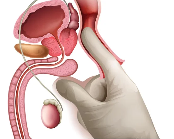
2. Insertion of Ureteroscope
A small, flexible or rigid tube with a camera (ureteroscope) is inserted through the urethra, bladder, and into the ureter.
3. Stone Visualization
Once the stone is located, its size and shape are examined.
4. Laser Fragmentation
A laser fiber (commonly Holmium:YAG) is used to break the stone into tiny pieces.
5. Removal or Natural Passage
Stone fragments are either removed with a basket tool or left to pass naturally through urine.
6. Stent Placement (Optional)
A ureteral stent may be placed temporarily to allow for smooth urine drainage and reduce swelling.
Postoperative Care & Recovery
Hospital Stay
Most patients can return home the same day.
Recovery Time
You can usually resume daily activities within 2–3 days, though complete recovery may take up to 2 weeks.
Side Effects
- Mild discomfort or burning during urination
- Temporary blood in the urine
- Frequent urination
- Stent Removal
If a stent is inserted, it’s typically removed within 5 to 14 days during a short outpatient procedure.
Benefits of Ureteroscopic Lithotripsy Surgery
- Minimally Invasive: No external incisions
- High Success Rate: Especially for mid-to-lower ureteral stones
Quick Recovery
- Can Treat Multiple Stones in One Session
- Fewer Complications Compared to Open Surgery
Conclusion
Ureteroscopic Lithotripsy Surgery is a proven and safe option for patients suffering from kidney or ureteral stones that are too large or painful to pass naturally. With minimal risks and high success rates, it has become the preferred surgical method for urologists around the globe.
If you’re experiencing persistent urinary pain, blood in urine, or recurring infections, it’s vital to consult a urologist for evaluation. Timely diagnosis and treatment — especially with advanced methods like ureteroscopic lithotripsy — can prevent complications and restore your health quickly.