


Diagnosis for Radiation oncology surgery is a critical pillar in the fight against cancer. It uses high-energy radiation to target and destroy cancerous cells. While the treatment itself is highly specialized, the diagnosis process for radiation oncology surgery is equally important. Accurate diagnosis ensures that patients receive the right treatment at the right time, maximizing effectiveness and minimizing harm. While the delivery of radiation is highly technical and specialized, the success of radiation oncology surgery begins long before the first treatment session. It starts with a comprehensive and accurate diagnostic process, which lays the groundwork for effective treatment planning. A misstep in diagnosis can lead to inappropriate treatment, unnecessary radiation exposure, and reduced survival outcomes.
The diagnostic phase involves a series of detailed evaluations including physical exams, imaging scans, biopsies, and laboratory tests all aimed at understanding the tumor’s size, location, type, and behavior. Only with a clear and complete picture of the disease can radiation oncology design a treatment plan that is both safe and highly targeted.
What is Radiation Oncology?
Radiation oncology is a medical specialty that uses ionizing radiation to treat cancer. Unlike surgical oncology, which removes tumors through surgery, or medical oncology, which uses chemotherapy, radiation oncology relies on precisely targeted radiation to shrink or eliminate tumors.
Radiation can be administered in various forms, including :-
-
External Beam Radiation Therapy (EBRT)
-
Brachytherapy (internal radiation)
-
Stereotactic Radiosurgery (SRS)
-
Proton Therapy
However, before any of these can begin, a comprehensive diagnosis and planning phase must be completed.
Why Is Diagnosis So Important in Radiation Oncology Surgery?
A detailed diagnosis is not only about identifying the presence of cancer; it is about understanding :-
-
The type and stage of cancer
-
Exact tumor location and size
-
Spread to nearby lymph nodes or organs
-
Patient’s overall health and response to prior treatments
This data forms the basis of a customized radiation therapy plan, reducing damage to surrounding healthy tissue and improving outcomes.
Key Diagnostic Procedures for Radiation Oncology Surgery
Below is a step-by-step guide to the essential diagnostic procedures in radiation oncology.
Physical Examination and Medical History
The process begins with a thorough physical examination and a detailed review of the patient’s medical history. This includes :-
-
Symptoms the patient is experiencing
-
Previous cancer diagnoses or treatments
-
Family history of cancer
-
Lifestyle factors like smoking, alcohol, and occupational hazards
Imaging Tests
Imaging is the backbone of radiation oncology diagnosis. It helps in visualizing the tumor’s size, shape, and exact location. Common imaging tests include :-
-
CT Scan (Computed Tomography) :- Provides detailed cross-sectional images of the body. Often used in simulation planning for radiation therapy.
-
MRI (Magnetic Resonance Imaging) :- Offers high-resolution images of soft tissues, useful for brain, spinal cord, or pelvic cancers.
-
PET Scan (Positron Emission Tomography) :- Highlights metabolic activity in tissues, helping distinguish between active cancer and scar tissue.
-
X-rays and Ultrasound :- Useful in some cancers like breast or abdominal tumors.
These imaging techniques are sometimes combined, like PET-CT scans, to provide comprehensive insights.
Biopsy and Pathology
While imaging shows the presence of a suspicious mass, a biopsy confirms the diagnosis. It involves the extraction of a small tissue sample from the suspected area for microscopic examination.
Types of biopsies include :-
-
Fine Needle Aspiration (FNA)
-
Core Needle Biopsy
-
Surgical Biopsy (incisional or excisional)
A pathologist analyzes the sample to determine :-
-
Type of cancer (e.g., carcinoma, sarcoma)
-
Grade of the tumor (aggressiveness)
-
Specific molecular markers or mutations
This is vital for targeted therapy decisions and radiation planning.
Blood Tests
Blood tests aren’t usually diagnostic for cancer directly but play an essential supportive role. These include:
-
Complete Blood Count (CBC) :- Checks for anemia or infection.
-
Tumor Markers :- Proteins like PSA (for prostate cancer), CA-125 (ovarian), or CEA (colon).
-
Liver and Kidney Function Tests :- Essential before initiating radiation, especially if chemotherapy is combined.
-
Electrolyte Panels and Coagulation Profiles :- Ensure the patient is fit for treatment.
Genetic and Molecular Testing
In certain cancers, especially breast, lung, and colorectal, genetic profiling is performed to :-
-
Identify mutations like EGFR, ALK, HER2, KRAS
-
Determine if targeted therapy or immunotherapy may work alongside radiation
-
Predict radio-sensitivity and possible side effects
Molecular diagnostics ensure the radiation therapy plan is personalized for each patient.
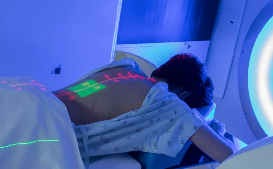
Simulation and Treatment Planning (Radiation Mapping)
This is a critical step unique to radiation oncology. Even before therapy begins, a simulation session is conducted :-
-
A CT scan simulation is done with the patient in the same position they’ll be during treatment.
-
Immobilization devices (like masks or body molds) are used for consistency.
-
The radiation oncologist marks the target area and organs at risk.
Using these details, a dosimetrist and medical physicist create a radiation plan with:
-
Exact beam angles
-
Radiation dose calculations
-
Safety margins to protect healthy tissues
Common Cancers Requiring Radiation Oncology Surgery
Radiation therapy plays a central role in treating many cancers, including :-
-
Brain tumors
-
Head and neck cancers
-
Breast cancer
-
Prostate cancer
-
Cervical and uterine cancer
-
Lung cancer
-
Rectal cancer
For some cancers, radiation is used before surgery to shrink the tumor (neoadjuvant), or after surgery to eliminate residual cells (adjuvant). In certain advanced stages, it is used palliatively to relieve symptoms.
The Role of a Multidisciplinary Team
Diagnosis and treatment planning for radiation oncology is not done in isolation. A team approach ensures comprehensive care. The team usually includes :-
-
Radiation Oncologist
-
Medical Oncologist
-
Surgical Oncologist
-
Radiologist
-
Pathologist
-
Dosimetrist and Medical Physicist
-
Oncology Nurse and Counselor
They work together to ensure the patient receives a personalized, safe, and effective radiation therapy plan.
Risks of Delayed or Inaccurate Diagnosis
An inaccurate or delayed diagnosis can result in :-
-
Treating the wrong area, leading to failure of therapy
-
Unnecessary radiation exposure to healthy tissue
-
Missed metastasis (spread of cancer)
-
Ineffective outcomes, especially in aggressive cancers
This is why early and precise diagnosis is critical to successful radiation oncology surgery.
Conclusion
Diagnosis in radiation oncology surgery is far more than identifying a tumor. It involves complex imaging, pathology, molecular biology, and simulation-based planning, all of which contribute to creating a highly personalized treatment roadmap.
Timely and accurate diagnosis can significantly improve treatment outcomes, minimize side effects, and offer the best chance of remission or cure.


