The brainstem is located at the junction of the midbrain, pons and medulla oblongata. It is the most inferior portion of the brain which acts as a center for many critical life processes. These include respiration, heart rhythm, blood pressure management and levels of consciousness. Due to the sensitive nature of this area, any abnormal growths such as tumors or lesions necessitate surgical procedures, which are often delicate in nature like brain stem surgery.
Brainstem Conditions is the removal of a brain stem tumor, which is commonly associated with other tumor-associated vascular malformations. There is a substantial need of mastery and understanding for diagnostic imaging, advanced surgical apparatus, microsurgical precision tools, along with hyper selective neurosurgical skill and mastery required for this form of surgery.
The approach of conducting this framework of surgery is often due to the absence of other viable forms of treatment. Successful outcomes from surgeries of this nature frequently lead to significant quality of life improvements, restoration of neurological functions, and many times becomes life saving.
Why Brainstem Conditions Is Performed
Brainstem Conditions is oftentimes a last resort. The following conditions are some of the most common reasons to operate on the brainstem:
- Life-threatening brainstem tumors such as gliomas and ependymomas.
- Certain vascular malformations such as cavernous malformations.
- Cysts or abscessed areas.
- Brainstem compression secondary to Chiari malformation.
- Severe trauma or hemorrhagic events.
- Uncontrollable trigeminal neuralgia or hemifacial spasm.
The surgery needs to be targeted toward controlling or ameliorating the growths or pressure while maintaining functional critical neuroanatomical structures. Minimally invasive and microsurgical approaches are standard practice in ensuring maximum safety and effectiveness.
Symptoms That May Indicate Need for Brainstem Conditions
Symptoms linked to Brainstem Conditions abnormalities tend to start subtle but worsen progressively. Since the Brainstem Conditions is involved in controlling bodily functions, even small lesions can have profound impact.
- Persistent headaches, in particular migraines, localized around the occipital area.
- Dizziness or balance problems.
- Diplopia or blurred vision.
- Hearing problems including loss of hearing or paroxysms of ringing in the ears (tinnitus).
- Dysphagia or swallowing difficulties.
- Facial numbness or weakness.
- Muscle weakness or paralysis of the limbs.
- Loss of coordination (ataxia).
- Difficulties in respiration.
- Altered states of consciousness or coma in severe cases.
Seeking a surgical assessment diagnosis rapidly for these brainstem disorders is imperative if one hopes to avoid significant irreversible neurological impairment or complications which can endanger one’s life.
Conditions Necessitating Brainstem Surgery
There are various medical conditions that may lead one to consider brainstem surgery. These conditions often present both benign and malignant growths.
Primary Causes for Brainstem Surgery:
- Brainstem Tumors:- Tumors such as gliomas, specifically ependymomas, medulloblastomas, and even metastatic lesions occur.
- Vascular Disorders:- Cavernous malformations, arteriovenous malformations (AVMs), or even aneurysms near the vertebrobasilar artery.
- Chiari Malformation: This is a congenital deformation which leads to brain tissues stretching into the spinal canal which in turn compresses the brainstem.
- Trauma or Hemorrhage:- This includes traumatic brain injury from either posterior fossa or internal bleeding.
- Infections and Abscesses:- Although quite rare, infection can cause the brainstem region to swell or form pus-filled cavities.
- Neurovascular Conflicts:- Conditions like trigeminal neuralgia or hemifacial spasm where a nerve is compressed causing abnormal movements.
Assessment: Identifying Disorders of the Brainstem
Evaluating and diagnosing a condition or disorders of the brainstem starts with a clinical workup including a physical examination, neuroimaging scans, and at times surgical exploration is necessary.
Diagnostic Methods Include:
- Neurological Examination:- Review and assess face, speech, coordination, and movement to check for muscle, and eye symmetries.
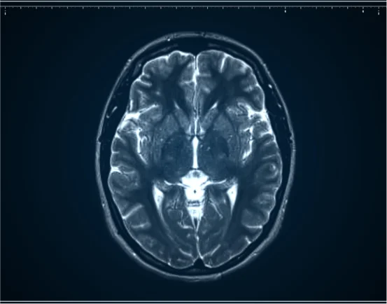
- Magnetic Resonance Imaging (MRI):- This diagnostic imaging method is effective in identifying and visualizing the stem and associated abnormalities.
- Magnetic Resonance Angiography (MRA) or CT Angiography (CTA):- These have also proven beneficial in identifying vascular abnormalities including aneurysms or AVMs.
- CT Scan:- While this test is often overlooked due to the precision and accuracy of an MRI, it can be lifesaving in emergencies or in cases of known internal bleeding.
Electrophysiological Studies:
Intraoperative monitoring is assessing the real-time active nerves of the brainstem during surgery.
Biopsy:
In some instances, a biopsy is critical in identifying the tumor type prior to treatment strategy formulation.
Treatment: Surgical and Non-Surgical Treatments for Disorders of the Brainstem
Surgical Treatment: Brainstem Conditions
Brainstem surgery is highly intricate and is done only in higher specialized centers with advanced neurosurgical capabilities. These centers apply modern techniques including:
- Microsurgery: Application of surgical telescopes/ microscopes and other precise tools
- Endoscopic Approaches: Access via the nose or mouth for minimal surgical intervention
- Neuronavigation Systems: Access through 3D brain mapping real time imaging
- Intraoperative Monitoring (IOM): During surgery to maintain motor and sensory functions.
Exact techniques used are tailored to the lesion’s location, size, and character.
Non-Surgical Treatment (When Possible):
- Radiation Treatment: Commonly adopted in cases of tumors that cannot be surgically removed
- Chemotherapy: For certain aggressive tumors, i.e gliomas
- Steroid Treatment: For management of cerebral edema
- Physical Rehabilitation: Active recovery after surgery and function maintenance
- Symptom Control: Relieving pain and speech/swallowing therapy as indicated
Risks and Recovery
As with any surgery, complications of the delicate anatomy of the brainstem, such as infection, hemorrhage, or nerve damage could occur. However, the combination of modern surgical technology and experienced brain tumor surgeons has provided good results for many patients.
Post-surgery recovery consists of:
- Monitoring in ICU for 24–72 hours
- Performing neurological evaluations
- Gradual rehabilitation: physiotherapy, occupational therapy, and speech therapy
- Follow-up MRIs for assessment of resorption and recurrence
Recovery duration may vary from a few weeks to several months, as it depends on the patient’s condition and the extent of the surgery performed.
Conclusion
Brainstem Conditions stands as one of the most complicated and remarkable advances in contemporary neurosurgery. At times, an equally impactful decision as undergoing these surgeries is the choice to undergo them. More often than not, it turns out to be the best—or only—hope for patients burdened with life-altering conditions such as tumors or vascular anomalies.
Advances in medical technology and surgical methods have made Brainstem Conditions safer and more effective than they have ever been. In the hands of seasoned neurosurgical teams, these surgeries perform wonders when it comes to dramatically enhancing survival rates, alleviating symptoms, and restoring lost quality of life.
If you or someone close to you has persistent neurological symptoms or a diagnosis of a Brainstem Conditions, seek out a certified neurosurgeon for a comprehensive neurological examination, and tailor-made treatment roadmap.