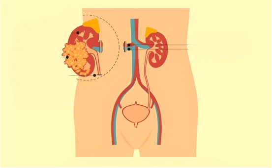
Pyeloplasty surgery is a type of surgery performed to treat a condition called ureteropelvic junction (UPJ) obstruction, which is the blockage of the urine flow from the kidney to the ureter. This blockage may lead to the swelling of the kidney, pain, infections, and even permanent damage to the kidney if not treated on time.
Why Is Diagnosis Important Before Pyeloplasty Surgery?
Understanding of the diagnosis for pyeloplasty surgery.
Correct diagnosis safeguards:
- Validation of UPJ obstruction
- Assessment of kidney function
- Detection of associated pathology
- Assessment of the need for surgery
- Avoidance of kidney damage
Considering that UPJ Obstruction symptoms could be absent or overlap with other conditions, diagnosis rests on sophisticated imaging modalities and function metrics.
Typical Symptoms That Aid In Diagnosis
Before any tests are ordered, many patients present symptoms that need further investigation. These are:
- Flank pain that is persistent or intermittent (more common to one side)
- Urinary tract infections (UTIs) recurrent in nature
- Hematuria (blood in urine)
- Nausea and vomiting, especially post drinking fluids
- Hydronephrosis (kidney swelling) which is often found incidentally
- Chronic renal insufficiency on lab tests
If these symptoms are longer lasting, a urologist may consider UPJ obstruction and would initiate imaging studies.
Imaging Studies For Pyeloplasty Surgery
The imaging studies for pyeloplasty surgery include several steps and tests to ascertain the presence of obstruction, determine its severity, and study how well the kidneys are functioning.
Ultrasound (USG) of the Kidney
Objective: Initial, atraumatic hydronephrosis indicator is USG of the kidney
- Reveals swelling in the renal pelvis
- Identifies certain anatomical pathologies
- Shall be used to assess post-operative progress
Ultrasound serves an integral role in identifying blockages however does not confirm functional obstruction.
Diuretic Renal Scintigraphy (Renal Scan/DTPA/MAG3 Scan)
- Objective: To evaluate function and drainage efficiency of the kidney.
- Measures how much each kidney works separately, including the percentage of function for each kidney.
- Uses radioactive dye and either a diuretic or Lasix.
This test is vital in deciding if surgery such as pyeloplasty is needed. Poor function but preserved drainage patterns typically necessitates surgery.
Intravenous Urography (IVU)
- Purpose: Imaging of the urinary system with x-rays post contrast injection of a radiologic dye.
- Visualizes parts of the body like the renal pelvis and ureter.
- Helps determine the severity of the obstruction.
Less common now, but still useful.
CT Urography
- Purpose: Imaging for complicated cases.
- Ultrasound offers limited value and renal scans fail to provide clarity; high-definition cross-sectional images.
- Stones, tumors, or structural abnormalities with especial attention on adults.
MRI Urography
- Purpose: Good for patients that should avoid radiation, like children and pregnant women.
- Gives details comparable to a CT untgraded scan. Good for repeat imaging.
- Additional diagnostic evaluation processes
Cystoscopy and Retrograde Pyelogram with anesthesia to visualize renal pelvis and ureter.
- Dye is injected into the ureter during cystoscopy.
- X-rays are taken to assess blockage.
- May be combined with temporary stenting for relief.
Causes of Ureteropelvic Junction (UPJ) Obstruction:
Understanding the underlying cause can impact diagnosis and surgical planning:
- Congenital obstruction (present at birth).
- Crossing blood vessels compressing the ureter.
- Kidney stones blocking the junction.
- Scar tissue or previous surgeries.
- Ureteral valves or strictures.
- Tumors or masses.
These can often be identifiable during imaging or surgical exploration.
When Is Pyeloplasty Surgery Recommended?
Surgery is usually advised when the diagnostic process reveals:
- Moderate to severe hydronephrosis.
- Significant functional obstruction on renal scan.
- Recurrent UTIs or pain.
- Declining kidney function.
- Non-resolving symptoms despite conservative management.
The standard procedure is Anderson-Hynes dismembered pyeloplasty which can be performed open, laparoscopic, or robotic depending on the case and hospital setup.
Benefits of Timely Diagnosis:
Accurate and timely diagnosis of UPJ obstruction with subsequent pyeloplasty surgery can:
- Preserve function of the kidney.
- Relieve painful symptoms.
- Prevent infections and complications.
- Improve overall quality of life.
Delayed diagnosis may lead to permanent damage or loss of function emphasizing the importance of prompt urological consultation.
Post-Diagnosis: What to Expect
After confirming a diagnosis and scheduling a surgery, patients might need to complete the following:
- Invasive procedures such as blood tests for renal function assessment
- Expire tests (urine) for possible infections
- Assessment of anesthesia
- Counseling before the operation
Imaging studies may need to be done 3-6 months post pyeloplasty to assess if the surgery has been successful.
Conclusion
The combination of ultrasound, renal scans, CT/MRI imaging, and sometimes direct endoscopic evaluations allow urologists to accurately determine the condition and formulate an effective management plan. Thus, early and accurate diagnosis is critical in managing UPJ obstruction undergo pyeloplasty surgically.
If you or a person you know struggles with flank pain, recurrent UTIs, or even kidney swelling, do not dismiss these signs – seek urological assessment to prevent advanced complications. Early assessment is crucial in preserving kidneys and lives.