Retinal detachment is a medical condition in which the retina, which is a photograph-sensitive tissue that resides at the posterior segment of the eye, is detached from its associated structures. This separation leads to dysfunction of the retina and in the long run will likely lead to vision loss. Seeking timely treatment for retinal detachment surgery is crucial for preserving vision.
Common Symptoms of Retinal Detachment:
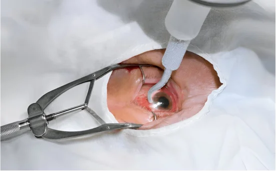
Understanding of the treatment for retinal detachment surgery
- Sudden onset of floaters or specks in vision
- Light flashes in one or both eyes
- Curtain-like shadow over the field of vision
Blurred or diminished central vision
Causes of Retinal Detachment:
- Aging (especially for individuals above 50 years of age)
- Severe ocular trauma
- Surgical intervention complications (for instance, cataract surgery)
- Diabetic retinopathy
- High myopia
Timely identification along with surgical intervention is vital for maintaining sight.
- Subtypes of Retinal Detachment:- Now, let’s discuss the three distinct forms of retinal detachment which may dictate different strategies for treatment and intervention:
- Rhegmatogenous Retinal Detachment:- Fluid accumulation beneath the retina as a result of fluid collection is rhegmatogenous retinal detachment, which is further classified by the break or tear in the retina.
- Tractional Retinal Detachment:- Surface scar tissue on the retina has the potential to pull away from the back part of the eye, resulting in tractional retinal detachment.
- Exudative Retinal Detachment:- The absence of a tear is characteristic of exudative retinal detachment. This form is commonly associated with tumor formation and other inflammatory processes.
Surgical management of retinal detachment requires a careful evaluation of the type and extent of retinal detachment, and may include one or more of the following treatment options:
Retinal Laser Surgery (Laser Photocoagulation)
Best for: Practiced for both pre-emptive and corrective measures, Ryggmatogenous tears or holes of the retina are best treated using laser photocoagulation.
Cryopexy (Freezing Therapy)
Best for: Classically indicated for small to moderate sized retinal tears, this form of therapy works through laser photocoagulation technique.
Procedure:
- An external freezing probe is applied over the eye directly above the tear. Cold temperatures induce a scar that aids in retinal adherence.
- Usually combined with: Scleral buckling or pneumatic retinopexy.
Pneumatic Retinopexy
Best for: Simple or small retinal detachments.
Procedure:
- A gas bubble is injected into the vitreous cavity.
- The patient is positioned so that the gas bubble pushes the retina toward the back of the eye.
- Sealing of the tear is performed using laser or cryopexy.
Recovery Note:
Patients are expected to maintain specific spatial head positions for several days to aid recovery.
Scleral Buckling Surgery
Best for: rhegmatogenous retinal detachment.
Procedure:
- A silicone band (scleral buckle) is attached vigorously over the white part of the eye (sclera).
- It gently pushes the eye wall inward relieving traction on the retina allowing reattachment.
Pros:
- Highly effective for complex and uncomplicated detachments.
- Usually performed under local anesthesia.
Vitrectomy
Best for: Complex or tractional retinal detachments.
Procedure:
- The vitreous gel exerting traction on the retina is surgically removed.
- The repositioned retina is secured with a gas bubble or silicone oil.
- Additional laser treatment or cryopexy is used to seal any remaining tears.
- Silicone oil must be removed at a later stage, whereas gas bubbles will dissipate naturally with time.
Recovery Following Retinal Detachment Surgery
The recovery process may differ according to the method of surgery performed, but general rules of thumb include:
Immediate Post-Surgery Guidelines:
- Avoid all forms of physical exertion
- Rest your eyes and remain in a relaxed state
- Apply prescribed eye drops to prevent inflammation and infection
- Maintain specific head positions if a gas bubble was administered
- Use an eye patch or shield as directed
Average Recovery Timelines:
Laser and cryopexy: A few days up to a week.
Vitrectomy or scleral buckle: Initial healing of 2 to 6 weeks; full recovery may take several months.
Potential Side Effects:
- Blurred or variable vision
- Slight discomfort in the eyes
- Transient floaters
- Redness and/or watering
It is essential to see your ophthalmologist for post-operative checks.
Outcome Metrics and Long-Term Projections
There is a high success rate treatment for retinal detachment surgery with retention of the detached retina. First surgery success falls within 80% to 90% range. However, the final visual prognosis is dependent on:
- The timeliness of detachment intervention
- Involvement of the macula (central retina)
- Patient eye health metrics and age
Treatment timing significantly impacts visual restoration outcomes by allowing early intervention to prevent lasting vision impairment.
Preventative Considerations for Retinal Health
While some retinal detachments are unavoidable, their likelihood can be minimized by doing the following:
- Scheduling regular eye checkups, particularly if one is myopic or has diabetes.
- Taking preventive measures to safeguard the eyes against trauma or injuries.
- Reporting any sudden vision changes (for example, flashes or floaters) without delay.
- Adhering to routine management of chronic conditions such as diabetes and hypertension.
Conclusion
Treatment for retinal detachment surgery is considered a medical emergency; however, vision can often be saved or significantly improved with accurate diagnosis and advanced surgical techniques. If you or someone you know suffers from sudden floaters, vision loss, or other concerning symptoms, do not wait—urgent evaluation and treatment by an ophthalmologist is essential. The likelihood of retaining vision after treatment for retinal detachment surgery is very high if intervention is done in a timely manner.