Hemispherotomy is a life changing neurosurgical procedure designed to treat severe and drug resistant epilepsy, particularly in children. Before undergoing this complex surgery, an accurate and comprehensive diagnosis is essential to determine whether the patient is an ideal candidate.
Hemispherotomy is a specialized neurosurgical procedure that involves functionally disconnecting one hemisphere (half) of the brain to stop the spread of seizure activity. This is particularly effective when seizures originate from one damaged or malformed hemisphere, a condition seen in disorders like Rasmussen’s encephalitis, hemimegalencephaly, and Sturge Weber syndrome.
What Is Hemispherotomy?
Hemispherotomy is a surgical procedure that involves disconnecting one cerebral hemisphere (either the right or the left side of the brain) to control epileptic seizures that originate from that hemisphere. Unlike hemispherectomy, which involves removing a portion of the brain, hemispherotomy is a disconnection procedure preserving the brain tissue but isolating its electrical activity.
This procedure is usually recommended in cases where seizures are frequent, debilitating, and do not respond to anti-epileptic medications. The goal of the surgery is to eliminate or drastically reduce the frequency of seizures, improving the patient’s quality of life.
When Is Hemispherotomy Considered?
Hemispherotomy is not a first-line treatment. It is considered only after all other treatment options, such as medications and less invasive procedures, have failed. Typically, patients considered for this surgery have the following conditions :-
-
Drug resistant epilepsy (intractable epilepsy)
-
Hemimegalencephaly (abnormal enlargement of one brain hemisphere)
-
Sturge Weber syndrome
-
Rasmussen’s encephalitis
-
Perinatal stroke or brain injury affecting one hemisphere
For such cases, where seizures significantly impair development, behavior, or day to day functioning, hemispherotomy offers a possible path to recovery. However, the procedure is only viable when comprehensive diagnostic testing confirms that seizure activity originates solely from one hemisphere.
Diagnosis of Hemispherotomy
Diagnosing a patient for hemispherotomy involves multiple evaluations. This process ensures that the surgical approach is appropriate and safe. Let’s look at the essential diagnostic steps :-
-
Detailed Medical History and Clinical Evaluation
The process starts with a comprehensive medical history. Doctors will collect detailed information about :-
-
Onset and frequency of seizures
-
Types of seizures experienced (e.g., tonic clonic, focal seizures)
-
Developmental milestones and any delays
-
Past treatments and their outcomes
-
Any associated neurological symptoms such as hemiparesis, speech delays, or cognitive impairments
A clinical neurological exam is also performed to assess motor functions, coordination, reflexes, and other neurological indicators. This helps identify any weakness or asymmetry in motor skills, which could point to unilateral brain dysfunction.
-
-
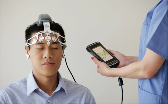
Video EEG Monitoring :- Electroencephalography (EEG) is one of the most important tools in epilepsy diagnosis. In the case of hemispherotomy, video EEG monitoring is often conducted over several days to capture seizure activity in real-time. This allows neurologists to :-
-
Determine the origin of seizures
-
Confirm that seizures consistently arise from one hemisphere
-
Rule out bilateral seizure activity (which would make hemispherotomy inappropriate)
The video component is crucial because it correlates electrical activity with physical seizure symptoms, helping map the brain-behavior relationship.
-
-
MRI (Magnetic Resonance Imaging) :- A high resolution MRI scan is essential for visualizing the brain’s anatomy. MRI helps identify structural brain abnormalities that may be causing epilepsy, such as :-
-
Cortical malformations
-
Tumors or lesions
-
Stroke or brain injury
-
Hemimegalencephaly or focal cortical dysplasia
The imaging must confirm that the abnormality is confined to a single hemisphere, supporting the case for hemispherotomy.
-
-
PET and SPECT Scans :- When MRI results are inconclusive or further functional imaging is needed, PET (Positron Emission Tomography) or SPECT (Single Photon Emission Computed Tomography) scans are used.
-
PET scans detect areas of decreased metabolic activity, often associated with epileptogenic zones.
-
SPECT scans performed during or just after a seizure (ictal SPECT) can show increased blood flow to the seizure focus.
These imaging studies add functional context to the anatomical findings of MRI and EEG.
-
-
Neuropsychological Assessment :- This assessment evaluates the patient’s cognitive, behavioral, and language abilities. The results help determine :-
-
The degree of functional impairment already present in the affected hemisphere
-
The risks and expected cognitive outcomes post-surgery
-
Whether the healthy hemisphere can support essential functions like speech and memory
If the dominant hemisphere (usually the left) is affected, the team must carefully evaluate potential post operative deficits.
-
-
Functional MRI (fMRI) and Wada Test (if required) :- In certain cases, a functional MRI or Wada test may be performed to assess language and memory functions in each hemisphere. This is especially important if the seizure focus is in the dominant hemisphere.
-
FMRI maps out brain activity during specific tasks, helping identify areas involved in speech, vision, and movement.
-
The Wada test involves injecting medication into one hemisphere at a time to assess the language and memory capabilities of each side.
These evaluations help surgeons plan the safest surgical strategy and avoid post-operative complications.
-
-
Multidisciplinary Team Discussion :- Once all the diagnostic information is collected, a multidisciplinary epilepsy team reviews the results. This team typically includes :-
-
Pediatric or adult neurologists
-
Epileptologists
-
Neurosurgeons
-
Neuropsychologists
-
Radiologists
The team works together to confirm that the patient is a suitable candidate for hemispherotomy and to design a personalized surgical plan.
-
Importance of Accurate Diagnosis Before Hemispherotomy
Hemispherotomy is a highly specialized surgery that can offer dramatic benefits but only when patients are carefully selected. An accurate and thorough diagnosis ensures :-
-
Proper identification of seizure origin
-
Minimization of surgical risks
-
Prediction of cognitive and motor outcomes
-
Selection of the correct surgical approach (lateral, vertical, or peri-insular)
Skipping or rushing any step in the diagnostic process can result in poor outcomes or unnecessary complications.
Conclusion
The diagnosis of hemispherotomy is a comprehensive and meticulous process that plays a pivotal role in ensuring the success of the surgery. From clinical evaluation and EEG monitoring to advanced imaging and neuropsychological testing, each step confirms whether the patient is an ideal candidate. While the surgery is complex, the benefits seizure freedom or significant reduction, improved quality of life, and better developmental prospects make the effort worthwhile for many patients and their families.