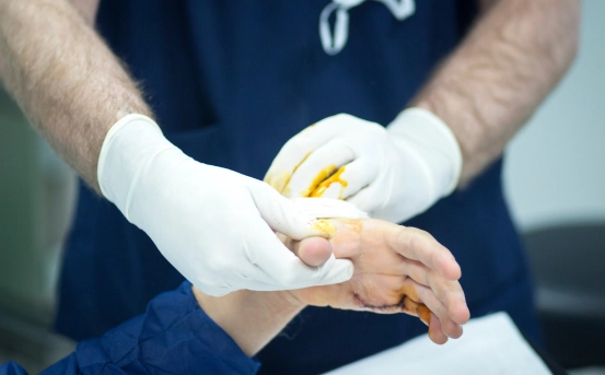
Splenectomy is a major surgical decision that is typically reserved for serious conditions where the spleen poses more harm than benefit. These may include traumatic splenic rupture, blood disorders like idiopathic thrombocytopenic purpura (ITP), certain cancers such as lymphoma, or infections that do not respond to conservative treatment. However, before proceeding with such a significant intervention, healthcare providers must conduct a thorough diagnostic process to determine whether spleen removal is truly necessary and beneficial for the patient.
The spleen is a small but essential organ located in the upper left part of the abdomen. It plays a crucial role in filtering blood, storing white blood cells and platelets, and fighting infections. However, under certain medical conditions, the spleen may need to be surgically removed a procedure known as splenectomy.
What is Splenectomy?
Splenectomy refers to the surgical removal of the spleen. While the spleen is not a vital organ and individuals can live without it, its removal may be necessary in specific cases such as traumatic injury, blood disorders, cancers, or spleen-related infections. The decision to remove the spleen is never taken lightly and involves a careful diagnostic process to evaluate whether the benefits of splenectomy outweigh the risks.
Why is Diagnosis Critical Before Splenectomy?
The spleen interacts with multiple systems in the body, especially the immune and circulatory systems. Removing it can make patients more susceptible to infections, particularly by encapsulated bacteria. Therefore, a thorough diagnosis ensures that splenectomy is justified, and no less invasive treatments would be more appropriate. Additionally, diagnosing the exact cause behind spleen related symptoms helps doctors determine whether total or partial splenectomy or even non surgical management is most suitable.
Common Indications Leading to Splenectomy
Several conditions can lead to the need for splenectomy. Each of these requires a distinct diagnostic pathway :-
-
Trauma or Injury :- Blunt abdominal trauma (like from a car accident) may rupture the spleen, necessitating emergency removal.
-
Hematological Disorders :- Conditions like idiopathic thrombocytopenic purpura (ITP), hereditary spherocytosis, thalassemia, or sickle cell disease may lead to an enlarged or overactive spleen.
-
Cancers :- Certain cancers such as lymphoma or leukemia may involve the spleen or cause it to enlarge.
-
Hypersplenism :- Overactivity of the spleen that causes the destruction of healthy blood cells.
-
Infections or Abscesses :- Severe infections or splenic abscesses may require removal of the organ.
Each of these indications is confirmed through a combination of clinical evaluation and diagnostic tests.
Clinical Evaluation: First Step in Diagnosis
Diagnosis begins with a detailed medical history and physical examination. Patients often present with symptoms such as :-
-
Pain or fullness in the upper left abdomen
-
Fatigue and weakness
-
Frequent infections
-
Easy bruising or bleeding
-
Anemia or low platelet count
On physical examination, doctors may detect splenomegaly (an enlarged spleen), tenderness, or signs of internal bleeding in trauma cases. These findings prompt further diagnostic imaging and laboratory tests to confirm the condition and the role of the spleen in causing symptoms.
Diagnosis of Splenectomy
Several non invasive imaging tests are used to assess the structure and function of the spleen :-
- Ultrasound :- Abdominal ultrasound is usually the first imaging test. It is safe, widely available, and helps detect splenomegaly, cysts, or masses in the spleen.
- CT Scan (Computed Tomography) :- CT scans provide a more detailed image of the spleen and surrounding organs. It is particularly useful in trauma cases to check for internal bleeding or rupture. Contrast-enhanced CT can also help assess blood flow and tissue damage.
- MRI (Magnetic Resonance Imaging) :- MRI is used in select cases to assess soft tissue detail and spleen abnormalities when further clarity is needed beyond ultrasound or CT findings.
- Nuclear Medicine Scans :- Spleen scintigraphy or radionuclide scanning helps assess spleen function and detect accessory spleens (additional spleen tissue) that may need removal.
Laboratory Tests Supporting Splenectomy Diagnosis
In addition to imaging, blood tests play a vital role in identifying the need for spleen removal :-
-
Complete Blood Count (CBC) :- Helps detect anemia, low white blood cell count, or low platelets.
-
Peripheral Blood Smear :- Offers insight into abnormal blood cells often seen in hereditary or autoimmune blood disorders.
-
Coagulation Profile :- Evaluates bleeding tendency, particularly in cases of hypersplenism.
-
Bone Marrow Aspiration/Biopsy :- Used when blood cancers are suspected to determine if the spleen is involved.
-
Autoimmune Tests :- Such as ANA, Coombs test, or reticulocyte count in cases of suspected autoimmune hemolytic anemia.
The combination of laboratory results and imaging findings guides the physician in understanding the extent of spleen involvement and the underlying condition.
Decision Making: When is Splenectomy Recommended?
The decision to proceed with a splenectomy is made after comprehensive diagnostic evaluation and multidisciplinary consultation. The following factors influence the recommendation :-
-
Severity of symptoms and the spleen’s impact on overall health
-
Failure of conservative treatments (e.g., medication for ITP or antibiotics for infections)
-
Risk of splenic rupture in trauma cases
-
Enlarged spleen causing pressure on nearby organs or leading to early destruction of blood cells
-
Cancer diagnosis where spleen removal helps stage the disease or relieve symptoms
If surgery is deemed necessary, pre operative vaccinations and planning are initiated to prepare for the post splenectomy phase, where infection risk is elevated.
Importance of Identifying Accessory Spleens
One crucial diagnostic aspect before splenectomy is the detection of accessory spleens. These are small nodules of splenic tissue separate from the main spleen. If not identified and removed, they can continue to contribute to disease symptoms especially in hematologic conditions like ITP. Imaging techniques like CT and radionuclide scans are useful for spotting accessory spleens.
Conclusion
Diagnosing the need for a splenectomy involves a meticulous and systematic approach. From clinical assessment and imaging studies to laboratory investigations and multidisciplinary evaluation, every step plays a role in ensuring that spleen removal is the right choice for the patient. Early diagnosis also helps in minimizing risks and improving long-term outcomes post surgery.