Introduction
Epidural hematoma is a serious medical condition that occurs when blood collects between the skull and the outer protective covering of the brain (dura mater). It is usually caused by a head injury or trauma and requires immediate medical attention. If not treated on time, it can lead to permanent brain damage or even become life-threatening. The most effective life-saving treatment is epidural hematoma surgery, also known as craniotomy for hematoma evacuation.
Understanding Epidural Hematoma
An epidural hematoma develops when an artery usually the middle meningeal artery ruptures after a skull fracture or severe head injury. The blood rapidly accumulates in the epidural space, creating pressure on the brain. This increased intracranial pressure can cause neurological damage within minutes to hours.
Key symptoms include:
- Severe headache
- Loss of consciousness (even brief)
- Vomiting
- Unequal pupils
- Weakness on one side of the body
- Confusion or drowsiness
If these symptoms appear after a head injury, the patient must be taken to the emergency room immediately.
Diagnosis Before Surgery
Doctors perform a combination of tests to confirm the presence of an epidural hematoma:
- CT Scan (Primary Test) :- A CT scan quickly reveals the location, size, and severity of the hematoma. It shows the classic “biconvex” shape of epidural bleeding.
- Neurological Examination :- This helps determine how the hematoma is affecting brain function.
- MRI (Occasionally Used) :- MRI is used when the CT scan results need additional clarity, although it’s less common in emergency situations.
Early diagnosis is essential because epidural hematomas can expand rapidly, leaving very little time for intervention.
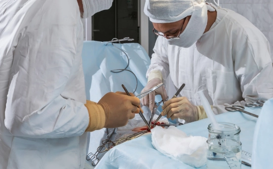
Treatment of Epidural Hematoma Surgery
The most effective and life-saving treatment for epidural hematoma is surgical evacuation, performed through a procedure called craniotomy.
- Craniotomy for Hematoma Evacuation :- This is the most common and preferred surgical method.
How it is done:
- The neurosurgeon makes an incision on the scalp.
- A small section of the skull (bone flap) is temporarily removed.
- The collected blood is carefully drained out.
- Bleeding vessels are sealed to prevent further bleeding.
- The bone flap is replaced and secured.
This procedure immediately reduces pressure on the brain and restores blood flow, improving the patient’s chances of recovery.
- Burr Hole Surgery (Used in Specific Cases) :- For smaller hematomas or patients who cannot undergo full craniotomy, a burr hole (small skull opening) may be used to drain the blood.
When Is Surgery Necessary?
Surgery is performed when:
- The hematoma is larger than 30 ml
- There is midline brain shift
- The patient shows worsening neurological symptoms
- High intracranial pressure is detected
- The patient has lost consciousness or has deteriorating GCS (Glasgow Coma Scale)
Recovery After Epidural Hematoma Surgery
Recovery depends on how quickly the patient received treatment and the severity of the brain injury.
- Immediate Post-Surgery Care :- Patients are monitored in the ICU for:
- Brain swelling
- Normal intracranial pressure
- Vital signs stability
- Risk of seizure
- Short-Term Recovery :- Patients may experience:
- Headache
- Mild confusion
- Fatigue
- Memory issues
These symptoms generally improve within days to weeks.
- Long-Term Recovery :- Most patients recover fully if surgery is done on time. Rehabilitation may include:
- Physiotherapy
- Speech therapy
- Cognitive therapy
Early physiotherapy helps restore muscle strength and balance.
- Potential Complications :- Though rare when treated on time, complications may include:
- Infection
- Seizures
- Brain swelling
- Rebleeding
- Neurological weakness
Regular follow-ups and medication can prevent most complications.
Importance of Timely Surgery
Epidural hematoma is one of the few neurosurgical emergencies where quick treatment almost guarantees excellent recovery. Studies show that patients treated within the “golden hour” recover significantly better.
Delays in treatment may lead to:
- Brain herniation
- Permanent disability
- Coma
- Death
This is why even a mild head injury followed by drowsiness or confusion should never be ignored.
How to Prevent Epidural Hematoma
While accidents cannot always be avoided, certain measures can reduce the risk:
- Always wear helmets while riding two-wheelers.
- Use seat belts while driving.
- Ensure children use proper head protection during sports.
- Install safety bars and railings at home for elderly individuals.
- Avoid high-risk activities when fatigued or intoxicated.
Conclusion
The treatment of epidural hematoma surgery is a critical intervention that saves lives by removing pressure on the brain and preventing irreversible damage. With advancements in neurosurgical technology, the success rate of this surgery has increased dramatically.