Introduction
Respiratory diseases are among the most common medical conditions worldwide, often requiring advanced imaging and interventional procedures for accurate diagnosis and effective treatment. Pulmonary interventions both vascular and non-vascular play a crucial role in identifying underlying abnormalities in the lungs, airways, and associated blood vessels. From detecting pulmonary embolism to managing lung lesions, diagnosis-driven interventions have transformed modern respiratory care. This blog explores the complete diagnostic pathway of vascular and non-vascular pulmonary interventions, types, imaging tools, indications, and clinical benefits, providing a clear understanding for patients, students, and healthcare professionals.
Diagnosis of Vascular and Non-Vascular Pulmonary Intervention
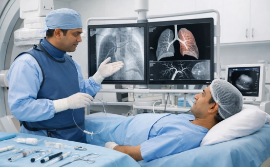
Pulmonary interventions involve minimally invasive procedures performed to diagnose or treat abnormalities affecting the lungs, bronchi, pleura, and pulmonary blood vessels. These interventions are generally guided by advanced imaging technologies such as CT, fluoroscopy, ultrasound, and digital subtraction angiography (DSA).
They are broadly classified into:
- Vascular Pulmonary Interventions :- These deal with pulmonary arteries, veins, and vascular complications, including:
- Pulmonary embolism
- Arteriovenous malformations (AVMs)
- Hemoptysis due to vascular causes
- Vascular stenosis
- Non-Vascular Pulmonary Interventions :- These focus on lung tissues, pleural space, and airways, including:
- Lung biopsies
- Drainage procedures
- Bronchial interventions
- Ablation for tumors
Accurate diagnosis helps determine whether the condition is vascular or non-vascular and guides appropriate treatment planning.
Importance of Diagnosis in Pulmonary Interventions
Proper diagnosis helps in:
- Differentiating between vascular and non-vascular causes of symptoms :- Shortness of breath, chest pain, hemoptysis, and cough can arise from multiple conditions; diagnostic interventions pinpoint the exact cause.
- Guiding targeted treatment :- For example, confirming pulmonary embolism ensures timely thrombolysis or thrombectomy, while lung nodules require biopsy-based evaluation.
- Reducing complications :- Precise imaging avoids unnecessary procedures and minimizes patient risks.
- Monitoring disease progression :- Interventions also help track response to ongoing treatments.
Diagnostic Tools Used in Pulmonary Interventions
Diagnosis of vascular and non-vascular pulmonary conditions relies on a combination of imaging methods and guided procedures:
- Computed Tomography (CT) Scan
CT imaging forms the backbone of pulmonary diagnosis.
- CT Pulmonary Angiography (CTPA) – crucial for detecting pulmonary embolism and vascular malformations.
- HRCT (High-Resolution CT) – helps evaluate interstitial lung diseases.
- Contrast-enhanced CT – highlights tumors, infections, and lymph nodes.
- Digital Subtraction Angiography (DSA)
Used in highly detailed evaluation of:
- Pulmonary artery stenosis
- Arteriovenous malformations
- Hemoptysis source
DSA is often followed by interventional procedures like embolization.
- Ultrasound
A helpful tool for diagnosing pleural effusion or guiding:
- Thoracentesis
- Pleural biopsies
- Chest drain insertion
It is safe, radiation-free, and ideal for bedside evaluation.
- Bronchoscopy
A key tool for non-vascular diagnosis.
Bronchoscopy allows direct visualization of airways and helps detect:
- Tumors
- Infections
- Obstructions
- Bleeding sources
It can also aid in taking biopsies.
- Fluoroscopy
Used during guided interventions such as:
- Lung biopsies
- Drainage procedures
- Bronchial interventions
Real-time imaging helps ensure safety and accuracy.
Diagnosis of Vascular Pulmonary Conditions
Vascular pulmonary interventions primarily deal with the blood vessels in the lungs. Diagnosis in these cases focuses on evaluating blood flow, obstructions, and abnormal vascular structures.
- Pulmonary Embolism (PE)
Diagnosis involves:
- CT Pulmonary Angiography (most preferred)
- D-dimer test
- Ventilation-perfusion (V/Q) scan
- Ultrasound of lower limbs (to detect DVT)
Interventions such as catheter-directed thrombolysis, mechanical thrombectomy, or IVC filter placement may follow depending on severity.
- Pulmonary Arteriovenous Malformations (AVMs) :- Abnormal connections between arteries and veins that may cause low oxygen levels.
Diagnosis includes:
- CT angiography
- DSA for detailed mapping
Treatment often involves embolization using coils or plugs.
- Hemoptysis (Bleeding in Lungs) :- When caused by vascular issues:
- Bronchial artery angiography
- CT scan to locate bleeding vessels
Diagnosis is critical so life-threatening bleeding can be managed through bronchial artery embolization (BAE).
- Pulmonary Hypertension & Vascular Stenosis :- Right heart catheterization, echocardiography, and CT angiography help determine vascular pressure and structural abnormalities.
Interventions include angioplasty and stenting if required.
Diagnosis of Non-Vascular Pulmonary Conditions
Non-vascular conditions affect the lung tissues, bronchi, alveoli, and pleura. Diagnosis aims to identify infections, tumors, inflammation, or structural abnormalities.
- Lung Nodules & Tumors
Diagnosis includes:
- CT scan to locate the lesion
- PET-CT to evaluate malignancy
- Image-guided lung biopsy for histopathological confirmation
Interventions such as radiofrequency ablation (RFA) or microwave ablation may follow for treatment.
- Pleural Effusion
Fluid in the pleural space is diagnosed using:
- Ultrasound
- CT scan
- Thoracentesis for fluid analysis
If large or recurrent, pleural catheter insertion or pleurodesis may be needed.
- Pneumothorax (Collapsed Lung)
Diagnosis includes chest X-ray and CT scan.
Interventions:
- Chest tube insertion
- Pigtail catheter drainage
- Lung Infections & Abscesses
Diagnosis may require:
- CT scan
- Sputum culture
- Guided drainage (if abscess)
- Airway Obstructions
Bronchoscopy plays a key role in diagnosis of:
- Foreign bodies
- Tumor blockages
- Mucus plug obstruction
Interventions include stenting, balloon dilation, or laser therapy depending on severity.
Benefits of Early Diagnosis in Pulmonary Interventions
-
- Prevents disease progression :- Early identification stops the condition from worsening.
- Minimizes complications :- Especially in vascular issues like pulmonary embolism.
- Enables targeted treatment :- Accurate diagnosis ensures correct procedures are performed.
- Improves survival and quality of life
- Supports long-term monitoring :- Interventions help track patient response effectively.
Conclusion
Diagnosis of vascular and non-vascular pulmonary intervention is a critical step in modern respiratory care. With advanced imaging, minimally invasive procedures, and skilled interventional radiologists, identifying lung diseases has become more efficient and accurate than ever. From detecting life-threatening pulmonary embolisms to evaluating lung nodules, precise diagnosis guides clinical decisions and ensures the best outcomes for patients.