Introduction
Adnexal tumors are growths that occur in the adnexa of the uterus, which includes the ovaries, fallopian tubes, and surrounding connective tissues. These tumors can be benign or malignant and are often discovered incidentally during routine pelvic examinations or imaging studies. Early and accurate diagnosis is crucial for effective treatment and improved patient outcomes. In this article, we explore the various methods used for the diagnosis of adnexal tumors, highlighting their importance and the latest advancements in medical imaging and laboratory testing.
Understanding Diagnosis of Adnexal Tumor
Adnexal tumors can originate from different tissues, including epithelial, stromal, and germ cells. While many adnexal masses are benign, such as ovarian cysts or fibrothecomas, some can be malignant, like ovarian carcinoma. Risk factors for developing adnexal tumors include age, family history of ovarian or breast cancer, genetic mutations such as BRCA1 and BRCA2, and hormonal imbalances.
Symptoms of adnexal tumors are often vague, including pelvic pain, bloating, abdominal distension, irregular menstrual cycles, and urinary frequency. Due to the nonspecific nature of these symptoms, clinical evaluation combined with diagnostic tools becomes critical for proper assessment.
Clinical Evaluation
The first step in diagnosing an adnexal tumor involves a thorough clinical evaluation. A detailed medical history helps identify risk factors and symptoms, while a pelvic examination allows the clinician to detect any palpable masses. However, physical examination alone cannot reliably differentiate between benign and malignant tumors, making imaging studies essential for further evaluation.
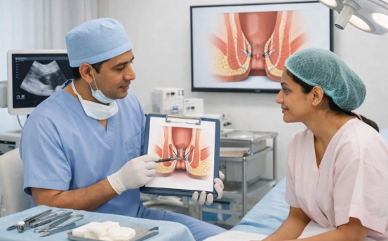
Imaging Techniques for Diagnosis
- Ultrasound : Transvaginal ultrasound is often the firstline imaging modality for evaluating adnexal masses. It provides highresolution images of the ovaries and surrounding structures, helping differentiate between solid and cystic lesions. Features such as septations, papillary projections, and irregular borders may raise suspicion for malignancy.
Doppler ultrasound can assess blood flow within the tumor. Malignant tumors often demonstrate increased vascularity compared to benign lesions, aiding in risk stratification.
- Computed Tomography (CT) Scan : CT scans are useful for assessing the extent of the disease, particularly in suspected malignant tumors. They provide detailed information about tumor size, location, involvement of adjacent organs, and the presence of metastases. CT imaging is also helpful in preoperative planning for surgical management.
- Magnetic Resonance Imaging (MRI) : MRI offers superior soft tissue contrast and is particularly valuable in characterizing complex adnexal masses. It can differentiate between different tissue types within the tumor, identify hemorrhagic or fatty components, and evaluate the tumor’s relationship with surrounding structures. MRI is often used when ultrasound findings are inconclusive.
- Positron Emission Tomography (PET) Scan : PET scans are primarily used in cases where malignancy is suspected or in the evaluation of recurrent tumors. PET imaging detects metabolic activity in tissues, helping to distinguish cancerous growths from benign lesions. It is often combined with CT (PET/CT) for precise anatomical localization.
Laboratory Tests
In addition to imaging, laboratory investigations play a vital role in the diagnosis of adnexal tumors. Tumor markers are substances produced by cancerous or noncancerous cells, which can be measured in blood tests. Commonly used tumor markers include
- CA125: Elevated in many ovarian cancers, though it can also rise in benign conditions like endometriosis or pelvic inflammatory disease.
- HE4 (Human Epididymis Protein 4): Often used alongside CA125 to improve diagnostic accuracy for ovarian malignancies.
- AFP (Alphafetoprotein) and βhCG: Helpful in detecting germ cell tumors.
- LDH (Lactate Dehydrogenase): Sometimes elevated in specific tumor subtypes.
While tumor markers provide valuable information, they are not definitive for diagnosis. They must be interpreted alongside imaging findings and clinical evaluation.
Histopathological Examination
Definitive diagnosis of an adnexal tumor requires histopathological examination. Tissue samples can be obtained through minimally invasive procedures like laparoscopy or imageguided biopsy. The pathological analysis determines the tumor type, grade, and potential malignancy, guiding appropriate treatment decisions.
Minimally Invasive Surgery
For many patients, laparoscopy is preferred for both diagnostic and therapeutic purposes. It allows direct visualization of the adnexa, removal of the tumor, and collection of samples for histopathology with minimal recovery time compared to open surgery.
Risk Assessment and Scoring Systems
Several scoring systems, such as the Risk of Malignancy Index (RMI), combine clinical, imaging, and laboratory parameters to estimate the likelihood of malignancy in adnexal tumors. These tools help clinicians make informed decisions regarding the need for surgery and referral to a gynecologic oncologist.
Importance of Early Diagnosis
Early detection of adnexal tumors, particularly malignant ones, significantly improves prognosis. Benign tumors may only require monitoring or elective removal, while malignant tumors benefit from timely surgical intervention and adjunct therapies like chemotherapy. Awareness of symptoms, routine gynecological checkups, and advanced diagnostic techniques contribute to better outcomes.
Conclusion
The diagnosis of adnexal tumors requires a multidisciplinary approach combining clinical evaluation, advanced imaging techniques, laboratory tests, and histopathological confirmation. Ultrasound remains the firstline tool, while MRI, CT, and PET scans provide additional information for complex cases. Tumor markers aid in risk stratification, and minimally invasive procedures allow definitive diagnosis and management.
Early detection and accurate diagnosis are key to improving patient outcomes, reducing complications, and providing targeted treatment. Regular gynecological examinations and timely investigation of pelvic symptoms are essential for anyone at risk. With ongoing advancements in medical imaging and molecular diagnostics, the future of adnexal tumor diagnosis is becoming increasingly precise and effective.