Introduction
Venous tumours are uncommon growths that develop from the veins or vascular tissues. They may be benign (non-cancerous) or malignant (cancerous), and their symptoms often resemble other vascular conditions. This makes accurate diagnosis extremely important. Proper evaluation ensures timely treatment, lowers the risk of complications, and helps specialists decide the most effective management plan.
What Are Venous Tumours?
Venous tumours are abnormal masses that originate from blood vessels, particularly veins. They may occur anywhere in the body, including the limbs, head and neck region, abdomen, or internal organs.
Common Types of Venous Tumours Include
- Hemangiomas benign vascular growths often found in children.
- Venous malformations congenital abnormalities that enlarge over time.
- Angiosarcomas are rare but aggressive malignant tumours arising from blood vessel walls.
- Hemangioendotheliomas tumours with intermediate behaviour (can be benign or malignant).
Because their appearance and symptoms overlap with other vascular issues, a structured diagnostic approach is essential to differentiate venous tumours from cysts, swelling, or lymphatic conditions.
Why Early Diagnosis Matters
Accurate and early diagnosis of venous tumours helps
- Reduce complications such as bleeding, infection, clots, or ulceration
- Identify the nature of the tumour benign, intermediate or malignant
- Guide treatment decisions such as surgery, medication, laser therapy, or interventional radiology
- Prevent progression especially in fast-growing or cancerous tumours
Delays in diagnosis can lead to tumour enlargement, pain, functional disability, or spread in case of malignant forms.
Diagnosis of Venous Tumours
- Clinical Examination :- A thorough physical examination is the first step.
What Doctors Look For
- Size, colour and texture of the lesion
- Presence of swelling, warmth, or tenderness
- Whether the mass is compressible
- Skin changes like bluish-purple discolouration
- Any functional impairment such as difficulty walking or moving a limb
Doctors may ask about symptoms like pain, heaviness, cosmetic concerns, bleeding history or sudden enlargement.
A detailed medical history helps determine whether the lesion is congenital or acquired and whether it may be associated with systemic conditions.
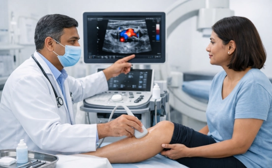
- Doppler Ultrasound :- Doppler Ultrasound is one of the most important tests used initially to assess suspected venous tumours. It is non-invasive, quick, and affordable.
What Doppler Ultrasound Reveals
- Blood flow characteristics within the tumour
- Presence of slow-flow or fast-flow vessels
- Extent of involvement (depth and diameter)
- Differentiation between venous and arterial components
Ultrasound also helps in identifying thrombus formation or calcifications inside the tumour.
- MRI Scan :- Magnetic Resonance Imaging (MRI) provides a detailed look at the tumour’s structure, depth, and relationship with surrounding tissues.
MRI Helps Determine
- Tumour boundaries
- Involvement of muscles, nerves, or bones
- Whether the tumour is aggressive or benign
- Internal vascular channels and tissue composition
MRI with contrast (MR angiography) offers enhanced visualisation of the vascular network and helps specialists plan surgical or interventional treatment.
- CT Scan or CT Angiography :- A CT scan is recommended when
- The tumour is located deep in the body
- Bone involvement is suspected
- Detailed vascular mapping is required
CT angiography helps detect feeding and draining veins, supporting treatment planning for embolisation or surgery.
- Venography :- Venography is used when detailed assessment of venous flow is needed. It involves injecting a special dye into the vein and capturing imaging with X-rays.
Venography Helps In
- Understanding abnormal venous pathways
- Detecting blockages or clots
- Evaluating surgical feasibility
Though less common today due to MRI advancements, it remains valuable in complex cases.
- Biops :- A biopsy may be required when
- Malignancy (like angiosarcoma) is suspected
- Imaging is inconclusive
- The tumour is rapidly growing or causing severe symptoms
Types of biopsies
- Fine needle aspiration cytology (FNAC)
- Core needle biopsy
- Excisional biopsy
Tissue samples are examined under a microscope to determine the tumour’s nature and grade.
- Blood Tests and Laboratory Investigations :- Although blood tests cannot directly diagnose venous tumours, they help assess
- Overall health
- Blood clotting status
- Signs of infection or inflammatory changes
- Tumour markers in rare cases of malignant vascular tumours
In some conditions, D-dimer levels may be elevated due to chronic clot formation within vascular malformations.
- Genetic and Molecular Testing :- Some venous malformations are linked to genetic changes such as TEK or PIK3CA gene mutations.
Genetic tests may be suggested when
- The tumour is congenital
- There are multiple vascular lesions
- Syndromic involvement is suspected
These tests help in predicting tumour behaviour and guiding targeted therapy.
- Differential Diagnosis :- Venous tumours can mimic
- Lymphatic malformations
- Arteriovenous malformations
- Soft tissue sarcomas
- Cysts or lipomas
- Varicose veins
- Deep vein thrombosis
Therefore, a combination of clinical assessment and imaging is crucial for accurate diagnosis.
- Multidisciplinary Evaluation :- Diagnosis and management of venous tumours often require a team of specialists, including
- Vascular surgeons
- Dermatologists
- Interventional radiologists
- Oncologists (for malignant cases)
- Plastic surgeons
Multidisciplinary review ensures comprehensive care and precise treatment planning.
Conclusion
The diagnosis of venous tumours involves a structured approach combining clinical examination, advanced imaging, laboratory testing, and sometimes biopsy. Early and accurate diagnosis plays a crucial role in determining the tumour type, preventing complications, and guiding the most effective treatment strategy.
Whether benign or malignant, detecting venous tumours at an early stage significantly improves outcomes and quality of life. If you notice unusual swelling, discolouration, or a vascular mass, seeking timely medical evaluation is essential.