Acoustic neuromas-also called vestibular schwannomas or, less often, acoustic tumors-are slow-growing, non-cancerous masses that form on the nerve linking the inner ear to the brain. Though they are not cancer, these lesions can gradually damage hearing and balance and diminish quality of life. How they feel, the range of treatments including advanced surgery, expected outcomes, and the steps of surgery and recovery. Whether you’ve just been told you have one or you are gathering facts for a loved one, the information here aims to help you move forward with clarity and confidence.
What is acoustic neuroma?
An acoustic neuroma springs from Schwann cells that cover the vestibular branch of the eighth cranial nerve, and it usually grows very slowly. Because the tumor sits at the base of the skull, it can press on nearby tissues, including the brainstem, and produce a mix of neurological signs. Experts label it vestibular schwannoma, and it accounts for roughly 8 percent of all tumors inside the skull.
Causes of Acoustic Neuromas
Experts have not pinned down a single cause of acoustic neuromas, yet several factors seem important:
- Genetic predisposition :- Inherited neurofibromatosis type 2 (NF2) sharply raises tumor odds.
- Environmental factors :- Researchers are still studying whether loud noise or cellphone radiation plays any role.
- Spontaneous mutation :- Most tumors arise sporadically with no obvious family history.
By tracking these risks, patients may spot warning signs early and adopt helpful lifestyle changes with their doctors.
Symptoms of Acoustic Neuromas
Early detection depends on noticing acoustic neuroma symptoms. Common clues include:
- Hearing loss :- Usually slow and confined to one ear.
- Tinnitus :- Persistent ringing or buzzing with no outside source.
- Balance issues :- Dizziness, unsteadiness, or sudden spinning.
- Facial numbness or weakness :- Tumor pressure on nearby nerves can cause tingling or reduced muscle control.
- Headaches and ear fullness :- Ongoing pressure or discomfort.
Seek prompt evaluation by an ENT or neurologist if two or more signs appear together.
Diagnosis and Evaluation
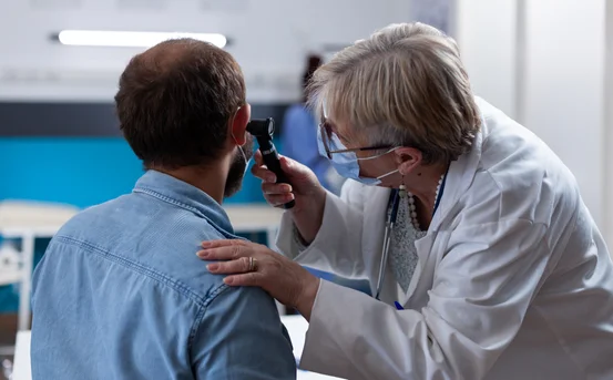
Diagnosing an acoustic neuroma typically involves a combination of symptom review, hearing tests, and imaging studies. A neurologist or ENT (ear-nose-throat) specialist will first take a medical history and perform a thorough ear and neurological exam. Since hearing loss is a common issue, an audiogram (hearing test) is usually ordered. This test measures hearing in each ear separately and can detect even subtle differences.
The definitive test for acoustic neuroma is magnetic resonance imaging (MRI) with contrast dye. An MRI scan produces detailed images of the brain and inner ear, allowing doctors to see even very small tumors (as small as 1–2 mm). If MRI is not possible (for example, due to a pacemaker), a CT scan with contrast might be used instead, although it is less sensitive for tiny tumors.
Treatment Options for Acoustic Neuroma
Once an acoustic neuroma is diagnosed, treatment decisions are based on tumor size, growth rate, patient age, health, and symptoms. The main options are observation (monitoring), surgery, and radiation therapy. In many cases, supportive care (like hearing aids and therapy) is also used to manage symptoms.
- Observation (Watchful Waiting): If the tumor is very small, not growing, and causing few or no symptoms, doctors may recommend simply monitoring it. This involves regular MRI scans (typically every 6–12 months) and hearing tests to ensure the tumor isn’t changing. Many acoustic neuromas grow so slowly that active intervention can be delayed or avoided, especially in older patients or those with other health issues.
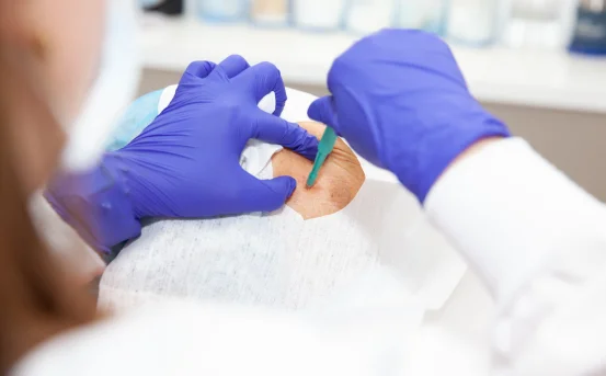
- Surgery (Acoustic Neuroma Surgery): Surgery is the primary treatment for larger or symptomatic tumors. An experienced neurosurgeon removes the tumor through the skull, using one of several approaches (translabyrinthine, retrosigmoid, or middle fossa craniotomy). The goal of surgery is to remove the tumor while preserving the facial nerve (which controls facial movement) and as much hearing as possible. However, complete removal isn’t always possible if the tumor is very close to critical brain structures. Surgery is done under general anesthesia; it may involve approaching the tumor “through the ear” or by making a small opening in the skull. After removal, patients may experience changes in hearing, balance, or facial function if those nerves were affected during the procedure. Complications of surgery can include cerebrospinal fluid leakage, hearing loss, facial weakness, tinnitus, balance problems, and very rarely stroke or infection. Surgery is often recommended when the tumor is large, growing, or causing significant symptoms, since removing it can relieve pressure on nerves and the brain.
- Radiation Therapy (Stereotactic Radiosurgery): This option uses focused radiation beams to stop the tumor from growing. Common techniques include Gamma Knife and CyberKnife radiosurgery. Unlike traditional radiation therapy, stereotactic radiosurgery delivers many small, precise doses of radiation to the tumor in one session (or sometimes a few sessions). It is often used for small to medium tumors (generally under 2.5–3 cm) or in patients who are not good candidates for open surgery. The goal is to “cook” the tumor so that it stops growing, while minimizing damage to surrounding tissue. This is a noninvasive procedure (no incisions), so recovery is usually quick. Patients often go home the same day. Side effects can include temporary headaches, fatigue, or scalp discomfort. Over time (weeks to years), the tumor usually stops growing; occasionally it may even shrink. Follow-up MRIs are needed to ensure the tumor stays stable.
- Supportive Care: Whether treated or observed, patients often benefit from therapies that address symptoms. For hearing loss, options include hearing aids or cochlear implants (for severe loss). Balance and dizziness can be helped by vestibular rehabilitation (balance therapy) and physical therapy. Occupational therapy can assist with any vision or coordination issues. These supportive measures improve quality of life but do not treat the tumor itself.
Your healthcare team will tailor the plan to your needs. For example, a young patient with a growing tumor might be steered to surgery, while an older patient with a tiny, slow-growing tumor might simply be observed.
Recovery and What to Expect
- After Surgery :- Patients typically stay in the hospital for about 3–5 days. Initially, they go to an intensive-care or recovery unit until stable, then move to a regular floor. Common immediate after-effects include headache, nausea, and fatigue, which are managed with pain medications. Most doctors encourage light activity (like walking) soon to help balance recovery. Patients will often need to rest at home for several weeks. Activity is gradually increased; for instance, short daily walks help rebuild strength. Heavy lifting, straining, or high-impact exercise should be avoided for at least 6 weeks. Patients may have stitches or staples at the incision site, which are usually removed in about 7–10 days. The wound care instructions (no swimming, sun exposure, etc.) follow standard post-craniotomy guidelines. Facial nerve function is closely watched. It’s common to have some temporary facial weakness, since the facial nerve runs near the tumor. This can cause partial inability to blink or smile at first, but usually improves over weeks to months. Eye care (like artificial tears) is important if the eye won’t close fully. Hearing on the operated side is often reduced or lost. Balance may be off for days to weeks, and physical therapy may be recommended to re-train the brain to balance normally.
- Timeline :- According to patient guides, many people start feeling better after the first month. By about 4–6 weeks after surgery most can resume non-strenuous work. It may take 3–6 months (or longer) to feel fully recovered. Full recovery means returning to normal daily activities, walking steadily, and having one’s usual energy level. Even after recovery, patients need periodic MRIs (often annually) for several years to ensure no tumor regrowth.
- After Radiosurgery :- Because this treatment is non-surgical, recovery is much quicker. Patients typically go home the same day. They may feel mild fatigue or headache, but most can resume normal activities within a day or two. There is no healing from an incision, so there are no wound care needs. Over the following weeks and months, symptoms like tinnitus or dizziness often improve as the tumor stops growing. Regular MRI scans will monitor the tumor every 6–12 months. Side effects of radiation can sometimes appear later (such as transient swelling of the tumor), but your doctors will check for and manage these during follow-ups.
Life Expectancy and Long-Term Outlook
Because acoustic neuromas are benign and slow-growing, life expectancy is usually normal with proper treatment or observation. Modern treatments are very effective at controlling tumor growth and preserving neurological function. In fact, the East Bay Brain & Spine group notes that “most patients with this condition have a normal life expectancy”.
Long-term outcomes depend largely on the tumor’s size at diagnosis and the treatment’s success. Early detection generally means simpler treatment (or even watchful waiting) and fewer complications. Even after surgery or radiosurgery, most patients continue to lead active, normal lives. The main lasting issues tend to be hearing loss on the affected side (often permanent) and the need for rehabilitation if balance was affected. In extremely rare cases where a large tumor is left untreated, it could press on the brainstem and be life-threatening. That is why regular follow-up is crucial.
In summary, with regular medical care and monitoring, people with acoustic neuroma usually enjoy a good quality of life and normal lifespan.
Next Steps and Support
If you or a loved one has symptoms suggestive of an acoustic neuroma – such as new hearing loss in one ear or persistent tinnitus – don’t wait. Early evaluation by an ENT specialist or neurosurgeon can make a real difference. Our clinic’s experts have extensive experience diagnosing and treating vestibular schwannomas. We offer comprehensive care, including advanced imaging and minimally invasive treatment options.
Contact us today to schedule an evaluation or second opinion. Our team will listen to your concerns, explain all available options (from monitoring to surgery or radiosurgery), and work with you to develop a personalized treatment plan. With timely care and expert support, most patients with acoustic neuroma can go on to lead healthy, active lives.