Urine flow blockage at the ureteropelvic junction (UPJ) where the ureter and the renal pelvis join is a common condition handled with a pyeloplasty. If diagnosed early, patients can avoid the severe swelling and pain in kidneys, along with permanent damage. Increasing the urine flow post surgery, with reconstruction and removal of obstruction, defines the goal of a pyeloplasty.
Understanding the causes for pyeloplasty surgery is important for diagnosis and treatment as well as overall patient awareness. We will explore factors that lead to such an operating.
Why is Pyeloplasty Surgery Performed?
Pyeloplasty is most often administered in cases of UP junction stricture or obstruction, which results in hydronephrosis. In the absence of surgical intervention, this blockage can progressively lead to deterioration of kidney function. The choice to undertake pyeloplasty is based on clinical evaluation of the obstruction severity, symptomatology, and renal impact.
Key Causes for Pyeloplasty Surgery
Congenital Ureteropelvic Junction (UPJ) Obstruction
A prevalent issue, particularly in children, is congenital UPJ obstruction. This condition reflects a narrowing of the ureter in moccasin form. It could be due to:
- Underdevelopment of muscular tissues of the UPJ region
- Overly abundant or malformed vasculature that would press the ureter and cross it
- Insufficient drainage due to immaturity of the kidney’s drainage system
Congenital UPJ obstruction can be identified during prenatal ultrasounds or right after birth. However, some children remain symptom-free until much later in childhood.
Acquired Ureteropelvic Junction Obstruction
This type of obstruction happens later in life, and it might be caused by quite a number of factors:
Scar Tissue Formation
Infections, trauma, or prior surgeries involving the kidney or ureter can lead to rupture and fibrosis, where active and passive scarring occurs at the junction.
Kidney Stones
Damaging or recurrent calculus in renal issues may form large and inflammatory disorders that lead to cancerous changes to the ureteropelvic junction, which narrows, gets clogged, or becomes inflamed.
Inflammation and Infection
Surgical or Instrumental Trauma
Surgery involving the urinary tract or the placement of stents or catheters may lead to unintentional injury to the ureter, resulting in post-operative scarring or strictures.
External Compression by Blood Vessels
In certain patients, additional renal arteries or veins may compress the ureter at the junction between the ureter and the renal pelvis. While these vessels represent normal anatomical variants of partial or complete obstruction, they justify corrective surgery, such as pyeloplasty.
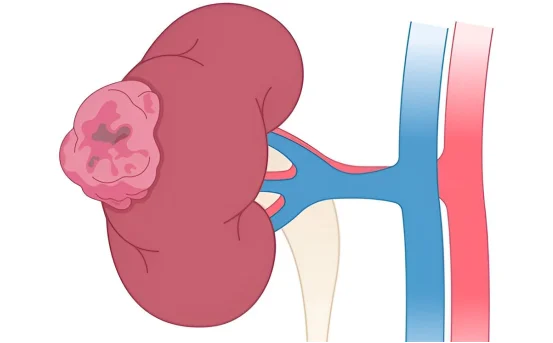
Tumors or Masses Near the UPJ
Tumors, whether benign or malignant, located within the kidney or adjacent soft tissues may compress the ureter and impede the flow of urine. Such obstruction may need to be treated with pyeloplasty in which the obstructive structure is bypassed or removed.
Ureteral Kinks or Twisting
The ureter may also be kinked or twisted in an abnormal fashion due to congenital factors or previous surgical interventions. Urine flow may be obstructed due to this anatomical alteration, and correction via pyeloplasty may be necessary.
Clinical Signs Which Could Signal Evaluation for Pyeloplasty
Some individuals may remain symptom-free, but many patients show signs of ureteropelvic junction obstruction, including:
- Flank or back pain, often occurring post fluid intake
- Recurrent urinary tract infections
- Blood in urine (hematuria)
- Nausea or vomiting, particularly after consuming fluids
- Palpable kidney mass in pediatric patients
- Imaging studies show hydronephrosis (swelling of the kidney)
Assessing Conditions Requiring Surgical Intervention of Pyeloplasty
An assessment should include:
- Ultrasound: Identify hydronephrosis or abnormal kidney size
- CT Urogram / MRI: Detailed imaging of the kidney and ureter.
- Renal Scan (MAG3 or DTPA): Evaluating renal function and drainage for that particular kidney.
- Voiding Cystourethrogram (VCUG): Evaluate for possible vesicoureteral reflux.
- Basic metabolic panel and urinalysis: Monitoring renal functions and infections.
These tests help determine the necessity of pyeloplasty surgery based on blockage and impairment assessed via imaging studies and test results.
Treatment: When is Pyeloplasty the Right Option?
Pyeloplasty Procedures Are Used in Situations Where:
- There is significant obstruction.
- The kidney is at risk of losing functional capability.
- There are recurrent infections or chronic pain.
Imaging studies indicate progressive hydronephrosis.
In relation to severity and the age of the patient, pyeloplasty can be conducted via:
- Traditional open surgery.
- Laparoscopic pyeloplasty.
- Robotic-assisted pyeloplasty.
All three methods seek to remove the obstructed segment and reconnect the healthy portions of the ureter and kidney.
Conclusion
Pyeloplasty surgery is a highly effective surgical intervention that restores normal urinary tract function and kidney preservation. It is most often necessary as a result of congenital UPJ obstruction; however, acquired causes such as scarring, stones, and even tumors are also frequent.
A prompt diagnosis and treating within timely manners of urinary obstruction is critical for the preservation of kidney function as well as reducing surgical interventions that can significantly improve quality of life. If you or a family member experiences recurrent flank pain, chronic urinary tract infections, or abnormal kidney scans, it is essential to speak with a urologist for an evaluation and determine if pyeloplasty is appropriate.