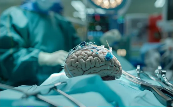
Craniotomy is a major brain surgery that can sound intimidating but for many patients, it’s a critical step toward treating brain tumors, bleeding in the brain, epilepsy, or even traumatic injuries. During this procedure, a portion of the skull is temporarily removed to allow surgeons access to the brain. After the necessary treatment is performed, the bone flap is replaced and secured.
Whether you or a loved one has been advised to undergo a craniotomy, understanding the procedure can help ease anxiety and prepare you for the journey ahead. From preparation to recovery, here’s a clear breakdown of what a craniotomy involves.
What is a Craniotomy?
A craniotomy is a surgical procedure where a piece of the skull called a bone flap is removed to allow direct access to the brain. The surgeon may perform this surgery to remove a tumor, drain bleeding, repair damaged blood vessels, relieve pressure, or perform a biopsy. After the brain surgery is completed, the bone flap is placed back in its original position and secured using plates or screws.
The size and location of the bone flap depend on the underlying medical condition and the area of the brain being treated.
Why is a Craniotomy Performed?
Craniotomy is done for several medical reasons, including:
- Brain tumors (benign or malignant)
- Traumatic brain injury
- Blood clots or hemorrhage
- Aneurysms or arteriovenous malformations (AVMs)
- Epilepsy surgery
- Brain infections or abscesses
- Pressure relief due to swelling (decompressive craniectomy)
Surgeons choose this procedure when direct access to the brain is necessary to diagnose or treat the condition effectively.
Craniotomy Surgery Procedure
Before surgery, patients undergo a detailed evaluation, which includes imaging tests like MRI or CT scans. These scans help the surgical team plan the safest and most precise route to the targeted area of the brain.
You may be asked to stop certain medications, avoid eating or drinking for a few hours before surgery, and follow other preoperative instructions. Your doctor will also explain potential risks, recovery expectations, and answer any questions you may have.
In some cases, patients may also meet with an anesthesiologist or neurologist before the surgery to assess readiness for general anesthesia or to plan for “awake brain surgery” if required.
During the Surgery
The craniotomy is typically performed under general anesthesia, meaning the patient is fully asleep and pain-free throughout the procedure. However, for tumors near areas that control speech or movement, an awake craniotomy may be performed. In this technique, the patient is kept awake and responsive during specific parts of the surgery to help surgeons avoid critical brain functions.
The procedure involves the following steps:
- Positioning and Incision :- The patient is positioned on the operating table, and the scalp is cleaned and shaved in the surgical area. The surgeon then makes a carefully planned incision in the scalp.
- Creating the Bone Flap :- A special surgical drill is used to create an opening in the skull. This bone flap is lifted and preserved for later replacement.
- Accessing the Brain :- Once the skull is open, the surgeon gently opens the dura (the membrane covering the brain) and begins the procedure such as removing a tumor or treating an aneurysm.
- Closure :- After the main part of the surgery is completed, the dura is closed, the bone flap is replaced and secured, and the scalp is stitched or stapled shut.
Depending on the complexity of the condition, the surgery may last several hours.
Recovery After Craniotomy
After surgery, the patient is monitored in an intensive care unit (ICU) for the first 24 to 48 hours. This ensures brain function, vital signs, and overall recovery are closely observed. Depending on how the surgery went and how the patient responds, they may remain in the hospital for several days.
Recovery at home includes rest, pain management, wound care, and gradual return to normal activities. Some patients may experience headaches, fatigue, dizziness, or changes in memory and speech. These usually improve with time and may be supported through physical or occupational therapy.
Follow-up appointments and imaging tests are scheduled to monitor healing and check for signs of recurrence or complications.
Risks and Complications
As with any major surgery, a craniotomy carries some risks. These can include:
- Infection
- Bleeding
- Swelling in the brain
- Seizures
- Blood clots
- Neurological deficits (speech, movement, vision, memory)
- Reactions to anesthesia
The risk level depends on the patient’s overall health, the specific brain condition, and the location of the surgery.
Conclusion
Craniotomy is a delicate and complex procedure but it’s also one that has saved and improved countless lives. Thanks to modern imaging, surgical techniques, and skilled neurosurgeons, outcomes are safer and more successful than ever before.
If you or a loved one is facing a craniotomy, having the right information and support system can make the journey more manageable. Open communication with your surgical team, following recovery guidelines, and staying informed will play a key role in a smooth recovery.