Apical Surgery encompasses procedures such as apicoectomy or root-end surgery. It is venturing into intricate dentistry reserved for situations when inflammation of the root cannot be resolved through conventional root canal treatment techniques. Different to regular dental work, this form of microsurgery has some degree of exactitude which must have in-depth knowledge and most importantly must start with precise diagnosis.
Prior to undertaking an apical surgery, critical understanding is important. Errors in the preliminary steps result in decisions that lead not only to unwanted surgical outcomes, but can also result infection within and around the mouth region thus affecting general health. Arrival at the decision “apical diagnosis” entails thorough clinical check-up and imaging examinations, alongside patient interactions.
Reasons Why Precise Diagnosis Is Mandatory In Apical Surgery
- Determining Root Cause Behind Relentless Infection
Some patients experience discomfort even after undergoing a successful root canal procedure. They may also develop swelling or show symptomatology suggestive of infection. In these cases accurate diagnosis help determine if problem exists within bone around root canal system, apex situated at lower extremity of the tooth’s root.
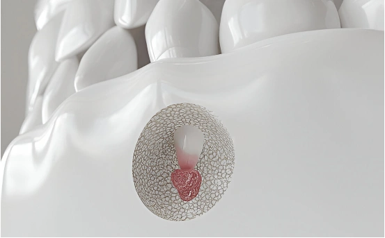
Understanding of diagnosis for apical surgery
Key factors that could justify performing apical surgery are:
- Periapical abscesses or cysts
- Fractured roots or untreated accessory canals
- Persistent granulomas
- Incomplete root canal fillings
Aside from detection and recognizing malignant conditions causing disproportionate pain which automatically directs towards performing diagnostically guided surgery such behavior will be accompanied without appropriate receiving adequate exploratory workup by searching out deeper lay transparent layers below hidden sub stratas would leads devoid devoided answer surrounding causes thus destitute solution reclamation remarks dispeers due absence’s identifiable groundwork under formulated guessworks .
- Avoiding Unnecessary Procedures
Accurate diagnosis aids in differentiating between cases that can be managed with conservative procedures like root canal retreatment, and those that require more surgical intervention. Apical surgery is an intricate procedure compared to standard root canal treatment, thus the clinician should ensure it is absolutely necessary.
With a cone-beam computed tomography (CBCT) scan, one can view 3D images of the region in question showing:
- Bone density loss
- Root canal anatomy
- Periapical pathology
- Sinusal involvement (in upper teeth{
Justifying the need for surgery becomes easier when conservative treatment options have been exhausted through thorough evaluation.
- Treatment Planning and Risk Assessment
Diagnosis not only validates the necessity for apical surgery but also assists in crafting a plan. During the diagnostic phase, factors such as tooth situs, bone topography, distance from vital structures (like the sinus cavity or nerves), general health status along with oral hygiene are evaluated.
For example:
- Surgery on a posterior tooth is higher risk owing to less access for viewing.
- Patients having systemic diseases such as diabetes or osteoporosis need special warnings.
- Appropriate diagnosis improves the customization of surgical strategy which enhances outcomes and shortens recovery time.
How Apical Surgery is Diagnosed: Important Steps
- Examination
The diagnostic steps start with a comprehensive clinical evaluation that involves:
- Assessing the tooth’s pain and swelling and its mobility
- Looking for sinus tracts, which are small pimple-like openings on gums
- Sensitivity examination by percussion and palpation
- Collection of medical and dental history from the patient
This physical assessment often points to whether or not there has been a failure in root canal therapy or additional surgical intervention is necessary.
- Radiographic Examination
Dental X-rays as well as CBCT scans are essential in diagnosing apical pathology.
Radiographs reveal:
- Periapical dark lesions:
- Root fractures or perforations;
- Previously missed canals;
- Signs of resorption
CBCT scans still provide three dimensional images which offer better assessment than traditional two dimensional x-rays particularly where complexity or uncertainty exists within cases.
- Vitality Tests Pulp
In some circumstances, the adjacent teeth are also subjected to vitality testing to determine if discomfort arises from the suspected tooth or its neighboring structures. This procedural step ensures that apical surgery isn’t performed on a healthy tooth situated adjacent to the one targeted.
- Microscopic Evaluation
Conventional imaging and the naked eye are insufficient to most endodontists who need a more refined approach like utilizing DOMs (Dental Operating Microscopes) that help detect vital inflammation, fractures or even dormant canals.
When is Apical Surgery the Best Option?
After a thorough assessment, apical surgery may be deemed necessary if:
- There exists an infection as a result of root canal treatment executed previously
- The area has intricate root structures that cannot be retreated
- Crown or post constructions sit over the tooth and does not allow for retreatment access
- Retreatments performed on the tooth have failed previously
- The bone structure surrounding the tooth is compromised thus requiring regenerative surgery
In such cases, apical surgery can be performed with the intent of preserving the natural dentition while avoiding extraction.
The Role of Endodontists in Diagnosis
A skilled specialist in diagnosing patients will go through great lengths in ensuring he performs thorough assessments before coming up with a plan to manage the issues identified surgically. Endodontists possess deep knowledge concerning roots, radiography interpretation and microsurgery which greatly benefits their patients because it eases receiving not only diagnostic but also treatment plans tailored specifically to them. They guide patients on possible complications during recovery timelines alongside steps towards optimal wellness post-surgery.
Conclusion
The treatment plan for apical surgery begins long before the patient takes a seat in the dental chair—accurate diagnosis sets the groundwork for everything. It’s crucial to understand that even the most meticulously performed surgical procedures may not succeed in the absence of proper diagnosis. Dental professionals use advanced imaging along with clinical evaluation and expert judgment to guarantee that apical surgery is performed only when absolutely indispensable and likely to yield optimal results.
As a patient suffering from unending dental discomfort or as an endodontist refining your next move, ensure you remember this one thing: Diagnosis isn’t the first step in this scenario. It happens to be, by all means, the cornerstone diverting factor which determines every outcome.