Introduction
Acute venous disorders are sudden conditions affecting the veins, often leading to pain, swelling, or circulatory complications that require timely medical attention. Early and accurate diagnosis not only helps control symptoms quickly but also prevents serious outcomes like pulmonary embolism or long-term venous insufficiency.
Understanding Acute Venous Disorders
Acute venous disorders refer to sudden, short-term conditions involving the veins. These include deep vein thrombosis (DVT), superficial thrombophlebitis, acute varicose vein complications, venous ulcers, and venous thromboembolism (VTE). These disorders affect blood flow within the veins and can rapidly worsen if not evaluated on time.
Common symptoms include
- Sudden leg swelling
- Pain or tenderness, especially in the calf
- Warmth or redness over the vein
- Visible enlarged veins
- Skin discoloration
Since these symptoms mimic other conditions like infections or muscle injuries, diagnosis requires a structured approach involving physical examination, imaging tests, and lab investigations.
Why Accurate Diagnosis Matters
Acute venous disorders are medical emergencies in many cases. For example, a blood clot in the leg (DVT) can travel to the lungs and cause pulmonary embolism, which is life-threatening. Similarly, untreated venous obstruction can lead to chronic swelling, skin damage, and long-lasting discomfort.
An accurate diagnosis helps
- Identify the exact location and severity of venous blockage
- Prevent clot migration
- Start timely anticoagulation or other therapies
- Reduce the risk of long-term complications
- Improve overall prognosis and patient safety
Diagnosis of Acute Venous Disorder
- Clinical Evaluation :- Diagnosis begins with a detailed medical history and physical examination. Doctors evaluate
Medical History
- Recent travel, prolonged sitting, or immobilization
- Previous episodes of DVT or thrombosis
- Family history of clotting disorders
- Hormonal therapy, pregnancy, or contraceptive use
- Recent surgery or trauma
- Cancer or chronic illness
These factors help determine the patient’s risk profile.
Physical Examination :- Doctors inspect the legs for
- Swelling differences between limbs
- Tenderness on calf compression
- Skin warmth or color changes
- Visibility of superficial veins
- Pitting edema
- Presence of varicosities
However, clinical signs alone are often not enough. Many cases of DVT occur with minimal external symptoms, requiring diagnostic tests for confirmation.
- Laboratory Tests for Diagnosis :- Laboratory markers help support clinical suspicion but are not always definitive.
D-dimer Test :- The D-dimer test is widely used as an initial screening tool. D-dimer is a protein fragment produced during clot breakdown. High levels suggest clotting activity inside the body.
- Useful for ruling out DVT or PE in low-risk patients
- Not diagnostic by itself can be elevated due to infections, pregnancy, surgery, or inflammation
A positive D-dimer typically leads to imaging tests.
Coagulation Profile :- This may include
- PT/INR
- aPTT
- Platelet count
These values help assess blood clotting ability and guide therapeutic decisions.
Thrombophilia Screening :- In patients with recurrent clots, young age, or family history, doctors may investigate inherited clotting disorders like
- Protein C and S deficiency
- Factor V Leiden mutation
- Antithrombin III deficiency
This helps plan long-term preventive strategies.
- Imaging Tests :- Imaging plays a crucial role in diagnosing acute venous disorders.
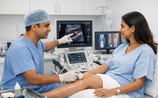
- Duplex Ultrasound :- Duplex venous ultrasound is the first and most reliable test for diagnosing DVT.
It evaluates
- Blood flow within veins
- Vein compressibility
- Presence of clots
- Degree of obstruction
Advantages include
- Non-invasive
- No radiation
- Quick results
- Widely available
Ultrasound is also used to detect superficial thrombophlebitis and assess venous insufficiency.
- CT Venography :- When ultrasound results are unclear or when clots are suspected in deeper veins (pelvis, abdomen), CT venography is recommended.
It provides:
- High-resolution imaging
- Accurate visualization of pelvic and abdominal veins
- Detection of large venous clots
CT venography is especially helpful in obese patients or those with complex anatomy.
- MR Venography :- MR Venography (MRV) is used when contrast exposure or radiation needs to be avoided.
Ideal for
- Pregnant patients
- Patients with kidney issues
- Suspected pelvic or abdominal venous obstruction
MRV provides detailed imaging of venous structures without radiation.
- Contrast Venography (Rare but Highly Accurate) :- Once considered the gold standard, contrast venography is now reserved for complex or unclear cases. It involves injecting contrast dye directly into the veins and taking X-ray images.
Used in
- Pre-surgical planning
- Cases where other imaging is inconclusive
Though highly accurate, its invasiveness limits its routine use.
- Additional Diagnostic Tools
- Ankle-Brachial Index (ABI) :- Used when arterial diseases coexist, helping differentiate venous from arterial circulation problems.
- Photoplethysmography (PPG) :- Evaluates venous refilling time to assess chronic venous insufficiency.
- Venous Pressure Studies :- Measure pressure changes within veins—used in specialized centers.
- Differential Diagnosis :- Venous disorder symptoms may resemble other conditions, making differentiation important
- Cellulitis
- Muscle tears or sprains
- Lymphedema
- Baker’s cyst
- Arterial diseases
A proper diagnostic approach eliminates these possibilities.
- Risk Stratification Tools :- Clinicians often use scoring systems to assess the probability of DVT.
Wells Score for DVT :- It scores pain, swelling, trauma, immobilization, cancer, and more
- High probability
- Moderate probability
- Low probability
This score helps decide whether a patient should undergo D-dimer testing or direct imaging.
- Importance of Early Detection :- Early diagnosis prevents severe complications such as
- Pulmonary Embolism (PE)
- Post-thrombotic syndrome
- Venous ulcers
- Chronic venous insufficiency
- Recurrent clots
Timely treatment leads to improved survival and better quality of life.
- When to See a Doctor :- Seek immediate medical evaluation if you notice
- Sudden leg swelling
- Pain or cramping
- Warm, red skin patches
- Prominent or hard superficial veins
- Unexplained shortness of breath
Early diagnosis saves lives, especially in conditions like DVT and VTE.
Conclusion
The diagnosis of acute venous disorders involves a combination of clinical assessment, laboratory tests, and advanced imaging techniques. With conditions like DVT and venous thromboembolism posing serious health risks, timely evaluation and accurate diagnosis are critical. Modern diagnostic tools such as duplex ultrasound, CT venography, and MRV have made detection faster and more precise. If you or someone you know experiences symptoms suggestive of a venous disorder, seeking prompt medical care can prevent life-threatening complications and ensure better outcomes.