Introduction
Atherosclerosis is a progressive disease in which the arteries become narrowed and hardened due to the accumulation of cholesterol, fats, and other substances. Over time, these deposits form plaques that obstruct blood flow, increasing the risk of heart attacks, strokes, and other cardiovascular complications. Early detection of atherosclerosis is crucial for preventing the progression of the disease and reducing the risk of severe complications. In this blog, we will explore the various diagnostic methods used to identify atherosclerosis and the importance of early diagnosis in managing the disease.
1. Physical Examination and Medical History
The first step in diagnosis of atherosclerosis often involves a thorough physical examination and review of the patient’s medical history. During the physical exam, the healthcare provider will check for signs of reduced blood flow in the arteries. Some of the things they may look for include:
-
High blood pressure (Hypertension) :- This is a common risk factor for atherosclerosis, and it can damage the walls of the arteries, making them more susceptible to plaque buildup.
-
Abnormal heart sounds :- A healthcare provider may listen for abnormal sounds, such as murmurs, which can indicate heart issues related to atherosclerosis.
-
Peripheral artery disease (PAD) :- In some cases, atherosclerosis may affect the arteries in the legs, causing poor circulation, pain, or visible signs of tissue damage.
In addition to the physical exam, the doctor will ask about the patient’s family history of cardiovascular diseases, lifestyle habits (such as smoking, diet, and exercise), and the presence of other risk factors like diabetes, obesity, or high cholesterol levels.
A family history of heart disease or stroke may raise suspicion of atherosclerosis, and lifestyle habits, such as smoking or a high-fat diet, can further indicate an increased risk of plaque formation.
2. Blood Tests
Blood tests are commonly used to assess the levels of substances in the blood that can contribute to the diagnosis of atherosclerosis. These tests are non-invasive and help identify underlying risk factors such as high cholesterol, diabetes, and inflammation. Some of the key blood tests used to diagnose atherosclerosis include:
1. Lipid Profile :- This test measures the levels of cholesterol and triglycerides in the blood. Key components of this test include:
- Total Cholesterol :- High total cholesterol levels are a major contributor to atherosclerosis.
- Low-Density Lipoprotein (LDL) :- Often referred to as “bad cholesterol,” high levels of LDL cholesterol can contribute to plaque buildup in the arteries.
- High-Density Lipoprotein (HDL) :- Known as “good cholesterol,” high levels of HDL cholesterol help remove excess cholesterol from the bloodstream and protect against atherosclerosis.
- Triglycerides :- High triglyceride levels are also associated with an increased risk of atherosclerosis.
2. Blood Glucose Test :- This test measures the level of glucose in the blood and is used to identify diabetes or insulin resistance, which are major risk factors for atherosclerosis.
3. High-Sensitivity C-Reactive Protein (hs-CRP) :- This test measures the level of inflammation in the body. Chronic inflammation plays a crucial role in the development and progression of atherosclerosis, making this test important for identifying at-risk individuals.
4. Homocysteine Levels :- Elevated levels of homocysteine, an amino acid, can damage the inner lining of the arteries and contribute to plaque buildup, increasing the risk of atherosclerosis.
While blood tests can’t definitively diagnose atherosclerosis, they provide important information about the underlying risk factors and can help doctors assess the likelihood of the disease.
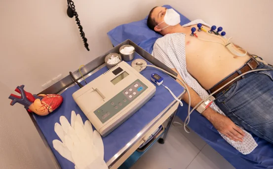
3. Electrocardiogram (ECG or EKG)
An electrocardiogram (ECG or EKG) is a non-invasive test that measures the electrical activity of the heart. While it does not directly diagnose atherosclerosis, it can provide valuable information about how well the heart is functioning and whether there are signs of heart damage due to reduced blood flow caused by atherosclerosis.
During an ECG, electrodes are placed on the chest, arms, and legs to detect the electrical signals produced by the heart. The resulting waveform provides information about the heart’s rhythm, heart rate, and conduction system. An abnormal ECG may indicate the presence of heart attacks, arrhythmias, or other heart-related issues associated with atherosclerosis.
4. Imaging Tests
Imaging tests are often used to directly visualize the presence of plaques in the arteries and assess the severity of atherosclerosis. These tests can detect narrowing or blockages in the arteries, helping doctors understand the extent of the disease and plan appropriate treatment. Some of the most commonly used imaging tests for diagnosing atherosclerosis include:
a. Ultrasound (Carotid Artery Ultrasound) :- A carotid artery ultrasound is a non-invasive imaging test used to evaluate the blood flow in the carotid arteries, which supply blood to the brain. The test uses sound waves to create images of the arteries and can detect the presence of plaque buildup. This test is particularly useful for identifying early signs of atherosclerosis, especially in individuals at risk of stroke.
If the ultrasound shows significant narrowing of the carotid arteries, it can indicate an increased risk of stroke due to decreased blood flow to the brain.
b. Ankle-Brachial Index (ABI) :- The ankle-brachial index (ABI) test is used to diagnose peripheral artery disease (PAD), which occurs when atherosclerosis affects the arteries in the legs. This test compares the blood pressure in the ankles with the blood pressure in the arms. A low ABI value indicates poor blood flow in the legs, which is often caused by atherosclerosis.
c. Coronary Angiography :- Coronary angiography is an invasive imaging procedure used to visualize the arteries of the heart and identify blockages or narrowing caused by atherosclerosis. During this procedure, a contrast dye is injected into the coronary arteries, and X-ray images are taken to detect any areas of plaque buildup.
This test is often performed when a patient is experiencing chest pain or shortness of breath, and there is a suspicion of coronary artery disease (CAD) due to atherosclerosis. Coronary angiography is considered the gold standard for diagnosing CAD.
d. CT Scan (Computed Tomography) :- A CT scan, or CT angiography, is a non-invasive imaging test that uses X-rays to create detailed cross-sectional images of the body. A CT scan can help detect the presence of atherosclerosis by identifying areas of plaque buildup in the arteries.
In some cases, a coronary calcium scan may be performed to evaluate the amount of calcium deposits in the coronary arteries. High levels of calcium in the arteries are indicative of atherosclerosis.
e. Magnetic Resonance Imaging (MRI) :- MRI is a non-invasive imaging test that uses magnetic fields and radio waves to produce detailed images of internal structures. An MRI can be used to assess the blood vessels and detect areas of plaque buildup, particularly in the heart and brain. In some cases, magnetic resonance angiography (MRA) can be used to evaluate blood flow and identify blockages caused by atherosclerosis.
5. Stress Tests
A stress test, also known as an exercise test, is used to evaluate how well the heart responds to physical activity. During a stress test, the patient is asked to walk on a treadmill or pedal a stationary bike while their heart rate, blood pressure, and ECG are monitored.
If the patient experiences chest pain, shortness of breath, or abnormal heart rhythms during the test, it may indicate that atherosclerosis has caused a significant reduction in blood flow to the heart. Stress tests are particularly useful for diagnosing coronary artery disease (CAD), which occurs when atherosclerosis affects the coronary arteries.
6. Other Diagnostic Tools
In addition to the above tests, several other diagnostic tools may be used to assess the presence and severity of atherosclerosis, including:
-
Magnetic Resonance Angiography (MRA) :- A non-invasive imaging test that uses magnetic fields and radio waves to produce detailed images of blood vessels.
-
Positron Emission Tomography (PET) :- A type of imaging that can detect inflammation in the arteries, which is an early sign of atherosclerosis.
Conclusion
Atherosclerosis is a progressive and potentially life-threatening condition that can lead to heart attacks, strokes, and other cardiovascular diseases. Early diagnosis is crucial to prevent complications and ensure effective treatment. Various diagnostic tools, including physical exams, blood tests, imaging tests, and stress tests, are used to detect atherosclerosis and assess the extent of arterial damage. By identifying the disease early, healthcare providers can help manage risk factors, slow disease progression, and improve patient outcomes.