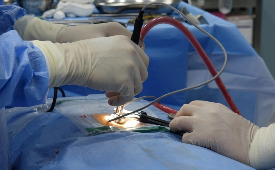
Introduction
Cervical fusion is not diagnosed as a condition itself but is recommended as a treatment after accurately diagnosing the underlying cervical spine problem causing pain, instability, or nerve compression. A proper and detailed diagnosis is essential to determine whether cervical fusion is necessary and to identify the exact spinal levels involved. The diagnostic process focuses on evaluating symptoms, identifying structural abnormalities in the cervical spine, and ruling out other causes of neck and nerve-related problems.
Diagnosis of Cervical Fusion
Clinical Evaluation and Medical History
The diagnostic process begins with a detailed medical history. The doctor asks about:
- Duration and severity of neck pain
- Radiation of pain to the shoulders, arms, or hands
- Presence of numbness, tingling, or weakness
- Difficulty with balance, coordination, or walking
- Previous neck injuries or trauma
- Past treatments such as medications, physiotherapy, or injections
- Impact of symptoms on daily activities and sleep
This information helps determine whether symptoms suggest nerve root compression, spinal cord involvement, or spinal instability—common reasons for cervical fusion.
Physical and Neurological Examination
A thorough physical and neurological examination is a key step in diagnosis. During this examination, the doctor evaluates:
- Neck range of motion and pain during movement
- Muscle strength in the arms and hands
- Reflexes to check nerve function
- Sensation to detect numbness or tingling
- Coordination and balance to assess spinal cord involvement
Signs such as muscle weakness, abnormal reflexes, or difficulty walking may indicate cervical myelopathy or nerve compression, strengthening the case for surgical intervention.
Imaging Tests
Imaging studies play a critical role in diagnosing conditions that may require cervical fusion.
- X-rays :- Cervical spine X-rays help identify:
- Loss of disc height
- Abnormal spinal alignment
- Bone spurs
- Signs of instability
Flexion and extension X-rays may be taken to assess abnormal movement between vertebrae.
- Magnetic Resonance Imaging (MRI) :- MRI is the most important imaging test for cervical spine diagnosis. It provides detailed images of:
- Intervertebral discs
- Spinal cord
- Nerve roots
- Soft tissues
MRI helps confirm disc herniation, spinal stenosis, cervical myelopathy, or nerve compression that may require cervical fusion.
- CT Scan :- A CT scan provides detailed images of bone structures and is useful for:
- Evaluating fractures
- Assessing bone spurs
- Planning surgical procedures
CT scans are often combined with MRI for a complete assessment.
Electrophysiological Tests
In some cases, nerve conduction studies (NCS) or electromyography (EMG) may be performed to evaluate nerve function. These tests help confirm whether symptoms are caused by cervical nerve compression or another neurological condition.
Diagnostic Injections
Selective nerve root blocks or epidural injections may be used diagnostically. If pain relief occurs after an injection, it helps identify the exact nerve level responsible for symptoms, assisting in surgical planning.
Ruling Out Other Conditions
Before recommending cervical fusion, doctors rule out conditions such as:
- Shoulder joint disorders
- Peripheral nerve injuries
- Muscle strain
- Inflammatory or infectious diseases
This ensures that surgery targets the correct cause of symptoms.
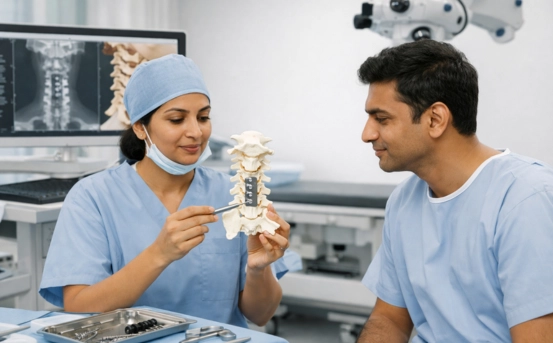
Determining the Need for Cervical Fusion
Cervical fusion is recommended only when:
- Symptoms persist despite conservative treatment
- Imaging confirms structural damage or instability
- Neurological deficits are present or worsening
- Spinal cord or nerve compression threatens function
The decision is made after correlating clinical symptoms with diagnostic findings.
Conclusion
The diagnosis leading to cervical fusion involves a comprehensive and systematic evaluation, including medical history, physical and neurological examination, advanced imaging studies, and supportive tests when required. Accurate diagnosis ensures that cervical fusion is performed only when truly necessary and that the correct spinal levels are treated.
Early and precise diagnosis not only improves surgical outcomes but also helps prevent long-term complications and neurological damage. Consulting an experienced spine specialist is essential for accurate evaluation, appropriate diagnosis, and successful treatment planning.