Introduction
Clubfoot (Congenital Talipes Equinovarus) is one of the most common congenital foot deformities found in newborns. Although the condition can look alarming to parents, early diagnosis and timely correction allow most children to grow up with normal, pain-free mobility. Understanding how is the diagnosis of clubfoot correction takes place and how this diagnosis influences the corrective approach is essential for caregivers seeking the best outcomes.
What Is Clubfoot?
Clubfoot is a structural deformity in which a baby’s foot is twisted out of shape or position. The affected foot typically turns downward and inward, making it appear as though the top of the foot is rotated toward the opposite leg. The condition may affect one or both feet. The diagnosis of clubfoot usually occurs at birth or even earlier during pregnancy, thanks to advancements in prenatal imaging. Once identified, correction can begin early often within the first few weeks of life.
Importance of Early Diagnosis of Clubfoot Correction
Early diagnosis of clubfoot is crucial for several reasons
- Supports early intervention, which leads to better correction outcomes.
- Reduces long-term disability, pain, and gait abnormalities.
- Improves success rates of non-surgical methods like the Ponseti technique.
- Allows parents to plan treatment, resources, and supportive care.
The earlier the diagnosis, the simpler and more effective the correction process.
Prenatal Diagnosis of Clubfoot
Many cases of clubfoot are now identified during the second-trimester ultrasound scan (18–22 weeks of pregnancy). Prenatal ultrasound can detect the abnormal foot positioning and reduced fetal limb movements.
How Prenatal Ultrasound Helps
- Confirms foot inversion or abnormal alignment
- Detects associated anomalies
- Assesses severity of deformity
- Helps parents prepare for postnatal treatment
However, prenatal diagnosis is not always 100% accurate. False positives may occur due to temporary fetal positioning. Therefore, post-birth evaluation remains essential.
Postnatal Diagnosis
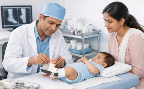
If clubfoot is suspected during pregnancy, or if foot deformity is visible at birth, a pediatric orthopedic surgeon performs a thorough physical examination.
Physical Examination
The doctor evaluates
- Foot position (downward, inward rotation)
- Tightness of tendons and ligaments
- Range of motion
- Muscle strength and development
- Foot length and shape
- Presence of creases on the inner border of the foot
- Knee and hip alignment
- Whether the deformity is flexible or rigid
The standardized evaluation helps determine the severity and best corrective method.
Pirani and Dimeglio Scoring Systems
To guide treatment, specialists often use rating scales
- Pirani Score :- This is the most widely used classification system. It scores six clinical signs
- Midfoot contracture (3 signs)
- Hindfoot contracture (3 signs)
Each is scored from 0 to 1, giving a total score of 0–6.
- 0–2: Mild deformity
- 2–4: Moderate deformity
- 4–6: Severe deformity
- Dimeglio Classification :- Evaluates four components
- Foot equinus (downward pointing)
- Varus (heel inward tilt)
- Adductus (forefoot inward)
- Cavus (high arch)
This system helps assess complexity and treatment requirements. These scoring systems allow doctors to track progress before, during, and after correction.
Imaging Tests for Clubfoot Diagnosis
Routine imaging is not always needed for typical clubfoot cases. However, certain situations may require additional imaging.
- X-rays :- Used when
- The deformity is rigid
- The diagnosis is unclear
- There is relapse after treatment
- Surgery is planned
X-rays help visualize bone alignment and joint orientation.
- Ultrasound (Postnatal) :- Useful for assessing
- Cartilage structure
- Joint positions in infants with flexible bones
- Atypical or syndromic clubfoot
- MRI or CT Scan :- Rarely needed, but beneficial in complex cases or when associated neuromuscular disorders are suspected.
Imaging supports clinical findings and enhances treatment planning when dealing with complex deformities.
Types of Clubfoot Diagnosed
Understanding the type of clubfoot is essential for planning corrections.
- Idiopathic Clubfoot :- The most common type, occurring in otherwise healthy infants.
Cause: multifactorial genetic and environmental. - Syndromic Clubfoot :- Occurs along with other congenital disorders like arthrogryposis or spina bifida.
This type is often more rigid and may require more extensive treatment. - Positional Clubfoot (Postural) :- Caused by improper fetal positioning.
It is flexible and typically resolves quickly with minimal intervention.
Treatment Planning After Diagnosis
Once clubfoot diagnosis is confirmed, treatment begins as early as possible preferably within 7–14 days of birth. The gold standard for correction is the Ponseti Method, a globally accepted non-surgical approach.
Ponseti Method Steps
- Gentle manipulation of the foot
- Weekly plaster casting to gradually correct deformity
- Percutaneous Achilles tenotomy (in most cases)
- Foot abduction brace for long-term maintenance
Success rate: 90–95% when done correctly and followed diligently.
If Ponseti treatment does not fully correct the deformity, surgical options may be considered, including tendon transfers or soft tissue release.
Red Flags During Diagnosis
Doctors check for signs that may indicate complex or atypical clubfoot
- Poor response to early casting
- Very stiff deformity
- Deep creases and shortened foot length
- Neurological abnormalities
- Abnormal reflexes or muscle weakness
These indicators help differentiate between idiopathic and syndromic clubfoot.
Parent Counseling After Diagnosis
A crucial part of clubfoot diagnosis is educating and preparing parents. Pediatric orthopedic teams explain
- Treatment steps
- Timeline and expected outcomes
- Brace-wearing importance
- Chances of recurrence
- Long-term follow-up needs
With proper guidance, parents become active partners in ensuring successful correction.
Why Early Diagnosis Leads to Better Correction
Early diagnosis allows
- Immediate initiation of the Ponseti method
- Avoidance of complex surgery
- Better foot shape and function
- Reduced recurrence rates
- Improved quality of life
Most children treated early lead fully normal and active lives.
Conclusion
The diagnosis of clubfoot is the first and most important step toward achieving complete correction. Through prenatal screening, postnatal physical examination, severity scoring, and selective imaging, specialists can accurately identify the condition and create a customized treatment plan. With timely intervention, especially using the Ponseti method, children with clubfoot can achieve normal, pain-free mobility and excellent long-term outcomes.