Introduction
Diagnosing disc herniation requires a combination of medical history, physical examination, and advanced imaging tests. Because the symptoms often resemble other spine-related problems, accurate diagnosis is essential to determine the right treatment plan. Below are the major diagnostic steps explained in detail.
Diagnosis of Disc Herniation
- Medical History Evaluation :- The first step in diagnosing disc herniation is a detailed medical history assessment. The doctor asks questions about the onset, duration, and pattern of your symptoms such as back pain, radiating leg pain, numbness, or weakness. They also enquire about past injuries, lifestyle habits like heavy lifting, long hours of sitting, and any previous spine problems. This discussion helps identify whether the symptoms are consistent with a herniated disc or caused by another spinal disorder.
- Physical Examination :- During the physical exam, the doctor checks your posture, spinal alignment, and pain points. They evaluate how well you can bend, twist, or move your spine. Strength testing is done to see if nerve compression is affecting muscle power. Reflexes are also tested slower or absent reflexes often indicate nerve involvement. The physical exam gives early clues about the affected spinal area (cervical, thoracic, or lumbar) and the severity of nerve compression.
- Neurological Assessment :- A neurological assessment checks the function of your nerves. The doctor inspects sensations like touch, temperature, and vibration to see if any nerve pathways are disrupted. They check for tingling, numbness, or muscle weakness by comparing both sides of the body. If a nerve root is pinched by the disc, it will show up as weakness or loss of feeling in specific areas. This test helps identify exactly which nerve is affected.
- Straight Leg Raise (SLR) Test :- The Straight Leg Raise test is commonly used to detect lumbar disc herniation. In this test, you lie on your back while the doctor gently lifts your leg while keeping the knee straight. If lifting the leg causes sharp, radiating pain down the leg, it suggests nerve irritation from a herniated disc. The test helps confirm whether the pain originates from the spine or another condition.
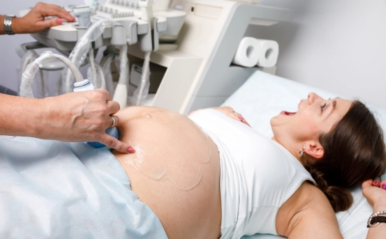
- X-Ray Imaging :- X-rays do not show herniated discs directly, but they are useful in ruling out other spine issues such as fractures, alignment problems, bone spurs, arthritis, or tumors. An X-ray gives a basic structural view of the spine and is usually the first imaging test ordered before more advanced scans.
- MRI (Magnetic Resonance Imaging) :- MRI is the most accurate and preferred imaging method for diagnosing disc herniation. It clearly shows the discs, nerves, spinal canal, and soft tissues. An MRI can detect the exact location, severity, and type of disc herniation, whether the disc is pressing on nerves, and whether inflammation or swelling is present. It helps your doctor plan whether you need medications, physiotherapy, injections, or surgery.
- CT Scan (Computed Tomography) :- A CT scan provides detailed cross-sectional images of the spine. It is often used when an MRI is not possible, such as for people with metal implants. CT scans offer clearer bone detail and help identify disc bulges, spinal canal narrowing, and other abnormalities. In some cases, CT is combined with a dye injection (CT myelogram) to show nerve compression more clearly.
- Electromyography (EMG) and Nerve Conduction Study (NCS) :- EMG and NCS tests measure electrical activity in your nerves and muscles. They are used when symptoms like numbness or weakness do not clearly point to a specific nerve root. EMG detects whether the nerve damage is from a herniated disc or another nerve disorder like neuropathy. NCS checks how fast electrical signals travel through your nerves. Together, these tests help confirm the level and severity of nerve compression.
- Discography (Rarely Used) :- Discography is performed only in complex or unclear cases. A special dye is injected into the suspected disc, followed by imaging. If injecting dye reproduces the same pain you normally feel, the disc is identified as the pain source. This method is mainly used before surgery when multiple discs appear abnormal and the surgeon needs to pinpoint the exact problematic disc.
- Differential Diagnosis :- Because disc herniation symptoms mimic many conditions like spinal stenosis, muscle strain, sciatica from other causes, infections, or joint problems the doctor also performs tests to rule out these conditions. Differential diagnosis ensures that the treatment focuses on the correct underlying issue and avoids unnecessary procedures.
Conclusion
Diagnosing disc herniation is a detailed process that involves history-taking, physical and neurological exams, and advanced imaging tests like MRI and CT scans. Early and accurate diagnosis helps prevent nerve damage, reduces pain faster, and guides the doctor in selecting the most effective treatment plan.