Introduction
Disc prolapse, commonly known as a slipped or herniated disc, is a condition that affects millions of people worldwide. It occurs when the soft inner core of an intervertebral disc pushes through the tougher outer layer, irritating nearby nerves and causing pain, numbness, or weakness in the affected area. Early diagnosis is crucial for effective management, preventing complications, and improving quality of life. This blog explores the key methods, tools, and considerations for the diagnosis of disc prolapse, helping patients and caregivers understand the process thoroughly.
Understanding Disc Prolapse
Before diving into diagnostic procedures, it is essential to understand what disc prolapse is. The spine is composed of vertebrae cushioned by intervertebral discs. These discs act as shock absorbers, allowing smooth movement of the spine. Over time, due to aging, trauma, or poor posture, the disc may weaken, and its inner gel-like nucleus pulposus may protrude through the outer fibrous ring.
Common symptoms of disc prolapse include
- Persistent back or neck pain
- Radiating pain in arms or legs (sciatica in lower back prolapse)
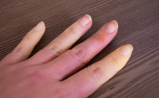
- Numbness or tingling sensations in extremities
- Muscle weakness
- Difficulty in movement or bending
Recognizing these symptoms early and consulting a healthcare provider is the first step toward proper diagnosis.
Step 1: Clinical Evaluation
The diagnostic journey begins with a detailed clinical evaluation. A physician will
- Review Medical History – Understanding previous injuries, lifestyle, occupation, and family history of spine disorders helps identify risk factors.
- Assess Symptoms – Doctors ask detailed questions about pain onset, duration, intensity, and any neurological symptoms like tingling or weakness.
- Physical Examination – This includes checking reflexes, muscle strength, range of motion, and sensory response. Tests like the Straight Leg Raise (SLR) can indicate nerve compression due to a prolapsed disc.
Clinical evaluation is vital because it helps determine the severity of the condition and guides further diagnostic tests.
Step 2: Imaging Studies
Imaging plays a crucial role in confirming disc prolapse and pinpointing its location. The most common imaging techniques include:
- Magnetic Resonance Imaging (MRI) :- MRI is the gold standard for disc prolapse diagnosis. It provides detailed images of soft tissues, including discs, spinal cord, and nerves. MRI helps determine
- The exact location of the prolapsed disc
- Degree of disc herniation
- Nerve compression
- Degenerative changes in adjacent vertebrae
MRI is non-invasive, safe, and highly effective, making it the preferred choice for most patients.
- Computed Tomography (CT) Scan :- A CT scan is often used when MRI is not suitable (for example, in patients with pacemakers). CT scans offer detailed images of bone structures and can show disc herniation indirectly by detecting spinal canal narrowing.
- X-rays :- While X-rays cannot directly show disc prolapse, they are useful in ruling out other causes of back pain, such as fractures, infections, or tumors. X-rays also help assess spinal alignment.
- Myelography :- In cases where MRI or CT scans are inconclusive, a myelogram may be performed. This involves injecting contrast dye into the spinal canal to highlight nerve compression on X-ray or CT imaging.
Step 3: Neurological Tests
To assess nerve function, doctors may recommend electrodiagnostic tests, including
- Electromyography (EMG) – Measures the electrical activity of muscles to detect nerve damage.
- Nerve Conduction Studies (NCS) – Evaluates the speed and strength of signals traveling through the nerves.
These tests are particularly useful in identifying which nerve roots are affected and assessing the severity of nerve involvement.
Step 4: Laboratory Tests
Though disc prolapse is primarily diagnosed via imaging and physical examination, laboratory tests may be ordered to rule out other conditions like infections or inflammatory diseases that mimic disc problems. Common tests include
- Complete blood count (CBC)
- Erythrocyte sedimentation rate (ESR)
- C-reactive protein (CRP)
These tests help ensure an accurate diagnosis by eliminating alternative causes of back or neck pain.
Step 5: Differential Diagnosis
Disc prolapse symptoms often overlap with other conditions. Accurate diagnosis requires differentiating from
- Spinal stenosis
- Osteoarthritis of the spine
- Muscle strain
- Tumors or infections of the spine
- Spondylolisthesis
A combination of clinical evaluation, imaging, and neurological tests helps confirm disc prolapse and rules out other causes.
Advanced Diagnostic Techniques
In complex cases, advanced diagnostic methods may be utilized
- Discography – Involves injecting a contrast dye into the disc to identify painful discs, often used before surgery.
- Functional MRI (fMRI) – Evaluates spinal cord activity and nerve function.
- Ultrasound Imaging – Sometimes used to assess soft tissue and muscle involvement around the spine.
These techniques are usually reserved for patients not responding to conventional diagnosis or treatment.
Importance of Early Diagnosis
Early diagnosis of disc prolapse is critical for
- Preventing nerve damage – Prolonged compression can cause permanent nerve injury.
- Effective pain management – Early intervention allows conservative treatments like physiotherapy, medications, and lifestyle modifications.
- Avoiding surgery – Mild cases often resolve with non-invasive measures if detected early.
- Improving mobility and quality of life – Prompt diagnosis prevents chronic disability and maintains daily function.
Common Treatment Approaches Post-Diagnosis
While this article focuses on diagnosis, it’s important to understand that accurate diagnosis directly influences treatment. Options include
- Conservative Management – Pain relief medications, anti-inflammatory drugs, physiotherapy, and lifestyle modifications.
- Minimally Invasive Procedures – Epidural steroid injections or nerve blocks for targeted pain relief.
- Surgical Intervention – Microdiscectomy or laminectomy may be recommended in severe cases where nerve compression persists.
Preventive Measures
Though not a substitute for medical diagnosis, maintaining spinal health can prevent disc problems
- Maintain proper posture while sitting or standing
- Exercise regularly to strengthen back muscles
- Avoid lifting heavy weights incorrectly
- Use ergonomic furniture and sleeping positions
Conclusion
The diagnosis of disc prolapse is a multi-step process involving clinical evaluation, imaging studies, neurological tests, and sometimes laboratory investigations. Early detection not only ensures timely management but also prevents long-term complications like chronic pain or permanent nerve damage. If you experience persistent back pain, numbness, or weakness, consulting a spine specialist is essential. With accurate diagnosis and appropriate care, most patients can manage symptoms effectively and return to their normal lifestyle.