Lymphoma is a type of cancer that originates in the lymphatic system, a crucial part of the body’s immune defense network. This system includes the lymph nodes, spleen, thymus gland, and bone marrow organs responsible for fighting infections and maintaining fluid balance. When cancer affects the lymphocytes, a type of white blood cell, it can lead to lymphoma. Because it can spread rapidly or grow slowly depending on the type, early and accurate diagnosis of lymphoma is essential for effective treatment and improved patient outcomes.
There are two primary categories of lymphoma: Hodgkin lymphoma (HL) and non Hodgkin lymphoma (NHL). Each behaves differently, requiring a tailored diagnostic and treatment approach. While some lymphomas may progress aggressively and need urgent intervention, others can be indolent and remain stable for years without immediate therapy. However, distinguishing between these forms is only possible through a series of diagnostic tests, imaging studies, and pathological evaluations.
What Is Lymphoma?
Lymphoma is a cancer that affects the lymphocytes, a type of white blood cell found in lymph nodes and other parts of the lymphatic system. There are two main types of lymphoma: Hodgkin lymphoma (HL) and non-Hodgkin lymphoma (NHL). Both types present similarly but differ in behavior, prognosis, and treatment options.
Because lymphoma can mimic other common illnesses or infections, accurate diagnosis often involves multiple steps and types of investigations.
When to Suspect Lymphoma?
Lymphoma can present with a wide range of signs and symptoms that are often subtle in the beginning. The most common symptom is painless swelling of the lymph nodes, typically in the neck, armpits, or groin. Other symptoms might include :-
-
Persistent fever
-
Night sweats
-
Unexplained weight loss
-
Fatigue
-
Shortness of breath or cough
-
Itching or skin rashes
When these symptoms persist without a clear cause, a doctor may suspect lymphoma and recommend further evaluation.
Initial Medical Evaluation
The diagnostic process usually starts with a physical examination and a thorough medical history. The physician will examine the size, location, and consistency of any swollen lymph nodes. They will also check for swelling in the spleen or liver and look for any other systemic signs.
If lymphoma is suspected, the next step typically involves imaging studies and blood tests to assess the extent and cause of the symptoms.
Diagnosis of Lymphoma
Imaging plays a key role in both detecting abnormal lymph nodes and staging lymphoma. Some commonly used imaging techniques include :-
- Chest X-ray :- A chest X-ray can help identify enlarged lymph nodes in the chest cavity or any associated lung abnormalities. It is often the first imaging test ordered.
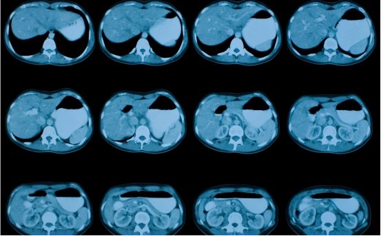
- CT Scan (Computed Tomography) :- CT scans provide detailed cross-sectional images of the body and help detect enlarged lymph nodes, tumors, or involvement of other organs. Contrast dye is often used to enhance clarity.
- PET-CT Scan (Positron Emission Tomography) :- A PET-CT scan is commonly used to detect areas of increased metabolic activity associated with cancer. This scan is especially useful in staging the disease and evaluating how aggressive it might be.
- MRI (Magnetic Resonance Imaging) :- MRI may be used in specific cases, especially if there is suspected involvement of the brain, spinal cord, or bones.
-
Blood Tests for Lymphoma :- While blood tests alone cannot confirm lymphoma, they provide essential information about overall health and how the body is functioning. Important blood tests include :-
-
Complete Blood Count (CBC) :- Helps detect anemia or abnormal white blood cell counts.
-
LDH (Lactate Dehydrogenase) :- Elevated levels may suggest tumor activity.
-
Erythrocyte Sedimentation Rate (ESR) :- An elevated ESR may indicate inflammation or infection.
-
Liver and Kidney Function Tests :- These assess how well organs are working, which is crucial before starting treatment.
-
-
Lymph Node Biopsy :- The definitive diagnosis of lymphoma requires a biopsy of the affected lymph node or tissue. This involves removing a portion of the lymph node, or sometimes the whole node, and examining it under a microscope.
Types of Biopsy
-
Excisional or Incisional Biopsy :- A surgeon removes an entire lymph node (excisional) or part of it (incisional) for detailed analysis. This is the most preferred method.
-
Core Needle Biopsy :- A large needle is used to remove a small cylinder of tissue.
-
Fine Needle Aspiration (FNA) :- This uses a thin needle to extract fluid and cells, though it may not always provide enough tissue for accurate subtyping.
The sample is sent to a pathologist for analysis, where they look for cancerous cells and determine the exact type of lymphoma.
-
-
Immunophenotyping and Flow Cytometry :- Once a biopsy is done, additional tests are performed to further classify the type of lymphoma. Immunophenotyping uses specific antibodies to detect certain markers on cancer cells. This helps distinguish between Hodgkin and non-Hodgkin lymphoma, and between various subtypes of NHL.
Flow cytometry is a specialized laboratory technique used to analyze the physical and chemical characteristics of cells in a sample. It helps determine the type of lymphocytes involved and whether they are behaving abnormally.
- Genetic and Molecular Testing :- Advanced tests like cytogenetics, FISH (Fluorescence In Situ Hybridization), and PCR (Polymerase Chain Reaction) are used to detect specific gene mutations or rearrangements. These findings can influence the prognosis and guide targeted therapy decisions.
- Bone Marrow Biopsy :- In certain cases, a bone marrow biopsy is required to check if lymphoma has spread to the bone marrow. This is done by extracting a sample from the hip bone using a needle. The presence of lymphoma cells in the bone marrow can affect staging and treatment planning.
-
Staging the Lymphoma :- After the diagnosis is confirmed, the next step is staging, which helps determine how far the cancer has spread. The Ann Arbor staging system is commonly used and ranges from Stage I (limited disease) to Stage IV (widespread involvement).
Staging involves a combination of imaging tests, bone marrow biopsy, and sometimes additional lab tests. Proper staging is essential to tailor the treatment plan to the individual’s needs.
How Long Does Diagnosis Take?
The entire diagnostic process may take anywhere from a few days to a couple of weeks, depending on the availability of biopsy results and imaging reports. While the waiting period can be stressful, it’s crucial to complete all evaluations for an accurate diagnosis and effective treatment plan.
Conclusion
Diagnosing lymphoma involves a series of tests and evaluations to accurately identify the type, stage, and spread of the disease. From initial physical exams and imaging to biopsies and molecular tests, each step plays a vital role in confirming the diagnosis and guiding treatment.