Introduction
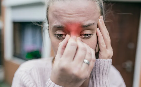
Spider veins are a common vascular condition characterized by thin, visible blood vessels that appear close to the surface of the skin, most often on the legs and face. Although spider veins are usually harmless and primarily a cosmetic concern, they can sometimes indicate underlying circulatory issues. Accurate diagnosis plays a crucial role in determining whether spider veins are purely superficial or associated with deeper venous problems that require medical attention.
The diagnostic process for spider veins is generally straightforward but comprehensive. It involves a detailed medical evaluation, physical examination, and, in some cases, specialized imaging tests. Proper diagnosis not only helps confirm the condition but also guides the most effective treatment plan and rules out serious vascular disorders.
Diagnosis of Spider Veins
Early diagnosis of spider veins is important for several reasons. First, it helps differentiate spider veins from other vascular conditions such as varicose veins or chronic venous insufficiency. Second, early evaluation allows doctors to identify potential risk factors, including poor circulation or vein valve dysfunction, before symptoms worsen. Finally, timely diagnosis ensures that patients receive appropriate advice on treatment options, lifestyle changes, and preventive care. While spider veins may initially appear as a cosmetic issue, delayed diagnosis can sometimes allow underlying venous problems to progress, leading to discomfort, swelling, or skin changes.
Medical History Evaluation
The diagnostic process begins with a detailed medical history. During this step, the doctor asks questions to understand the patient’s symptoms, lifestyle, and overall health. Key areas of discussion include
- When the spider veins first appeared
- Whether they are increasing in number or size
- Presence of symptoms such as pain, itching, burning, heaviness, or swelling
- Family history of vein disorders
- Occupational habits, especially prolonged standing or sitting
- History of pregnancy, hormonal changes, or use of hormonal medications
- Previous vein treatments or surgeries
This information helps the physician assess potential contributing factors and determine whether further diagnostic testing is needed.
Physical Examination
A thorough physical examination is a core component of diagnosing spider veins. The doctor visually inspects the affected areas, typically the legs or face, under good lighting. The examination may be performed while the patient is standing, as this position makes vein abnormalities more visible due to gravity.
During the physical exam, the doctor assesses
- Color, size, and distribution of visible veins
- Skin texture and temperature
- Signs of inflammation, discoloration, or skin thinning
- Presence of swelling or tenderness
- Any associated vein abnormalities beneath the skin
The doctor may gently press on the skin to evaluate blood flow and vein refill patterns. This hands-on assessment helps distinguish spider veins from other vascular or dermatological conditions.
Assessment of Symptoms
Although spider veins are often asymptomatic, some patients experience mild discomfort. Evaluating symptoms is an important part of diagnosis. The doctor will ask whether the patient experiences
- Aching or burning sensations
- Leg heaviness, especially after standing
- Mild swelling around the ankles
- Itching or skin irritation near visible veins
- Fatigue in the legs
Symptom assessment helps determine whether spider veins are associated with circulatory stress and whether additional diagnostic tests are required.
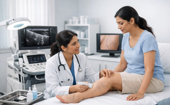
Use of Diagnostic Imaging
In cases where spider veins are extensive, symptomatic, or suspected to be linked to deeper venous issues, imaging studies may be recommended. These tests provide valuable insight into blood flow and vein function.
- Doppler Ultrasound :- Doppler ultrasound is the most commonly used imaging test in diagnosing vein-related conditions. It is non-invasive, painless, and does not involve radiation. This test allows doctors to evaluate
- Blood flow direction
- Vein valve function
- Presence of venous reflux
- Any blockages or abnormalities in deeper veins
Ultrasound helps confirm that spider veins are not associated with more serious venous disorders and ensures safe planning of treatment.
Differential Diagnosis
An important aspect of diagnosing spider veins is ruling out other conditions that may resemble or coexist with them. The doctor carefully distinguishes spider veins from
- Varicose veins
- Chronic venous insufficiency
- Capillary malformations
- Skin discoloration due to other medical conditions
- Inflammatory or autoimmune skin disorders
Accurate differentiation ensures that patients receive the correct diagnosis and appropriate care.
Evaluation of Risk Factors
Doctors also evaluate risk factors that may contribute to the development or progression of spider veins. These include
- Age-related changes in blood vessels
- Hormonal fluctuations
- Genetic predisposition
- Obesity or excess body weight
- Sedentary lifestyle
- Prolonged standing or sitting
Understanding these factors helps doctors provide personalized advice for prevention and long-term management.
Conclusion
The diagnosis of spider veins is a comprehensive process that goes beyond visual inspection. It involves a careful review of medical history, detailed physical examination, symptom assessment, and, when necessary, advanced imaging studies. Accurate diagnosis ensures that spider veins are correctly identified, underlying venous conditions are ruled out, and the most appropriate treatment strategy is chosen.
Early and thorough diagnosis not only improves treatment outcomes but also helps prevent potential complications related to circulation. By consulting a qualified healthcare professional and undergoing proper diagnostic evaluation, patients can gain clarity about their condition and take informed steps toward healthier veins and improved quality of life.