Introduction
Subarachnoid hemorrhage (SAH) is a life-threatening condition caused by bleeding into the space surrounding the brain, known as the subarachnoid space. It usually results from a ruptured cerebral aneurysm or head trauma. Early diagnosis is critical because within minutes, pressure inside the skull rises and can lead to permanent brain damage or death. Timely detection not only improves survival but also helps doctors determine whether emergency surgery or endovascular treatment is needed.
What Is a Subarachnoid Hemorrhage?
A subarachnoid hemorrhage occurs when blood leaks into the gap between the brain and the surrounding membranes. This space contains cerebrospinal fluid that helps cushion the brain. When blood fills this area, it irritates brain tissues, increases pressure, and disrupts normal blood circulation.
Common causes include
- Ruptured brain aneurysm (most common)
- Arteriovenous malformation (AVM)
- Traumatic head injury
- Bleeding disorders or blood thinners
- Rare genetic vascular disorders
Because it progresses rapidly, SAH is considered a neurological emergency. Quick and accurate diagnosis is essential to prevent rebleeding, stroke, and other complications.
Diagnosis of Subarachnoid Hemorrhage Surgery
Recognizing the warning signs helps prompt an immediate medical evaluation. Patients often describe the “worst headache of their life,” which begins suddenly.
Major symptoms include
- Sudden, intense headache
- Neck stiffness
- Nausea and vomiting
- Blurred or double vision
- Loss of consciousness
- Sensitivity to light
- Seizures
- Confusion or irritability
Anyone showing these symptoms should be taken to an emergency department immediately. Early suspicion is the first step toward an accurate diagnosis.
How Subarachnoid Hemorrhage Is Diagnosed
Diagnosing SAH involves a combination of clinical examination and advanced imaging tests. Doctors use a structured approach to confirm bleeding, locate the source, and plan further treatment especially if surgery or endovascular therapy is required.
- Physical and Neurological Examination :- Upon arrival, doctors conduct a quick but detailed neurological assessment. They check
- Pupil size and response to light
- Level of consciousness
- Neck stiffness
- Muscle strength and coordination
- Vital signs such as blood pressure
This helps determine how severe the hemorrhage might be and whether the patient is stable enough for imaging tests.
- CT Scan of the Brain: The First-Line Diagnostic Test :- A non-contrast CT scan is the fastest and most accurate method for detecting SAH in the first hours after symptom onset. In many cases, it can confirm the presence of blood with almost 100% accuracy within the first 6 hours.
Why CT scan is crucial
- Quick and widely available
- Identifies bleeding location
- Helps assess hydrocephalus (fluid buildup)
- Assists in predicting severity
If blood is found on the CT scan, the next step is to identify the cause—usually an aneurysm or vascular abnormality.
- CT Angiography (CTA) :- If a CT scan confirms SAH, doctors perform CT angiography to visualize the brain’s blood vessels.
CTA helps detect
- Aneurysms
- Arteriovenous malformations (AVMs)
- Vessel spasms
- Vascular injuries
CTA guides surgeons in planning either surgical clipping or endovascular coiling, depending on the aneurysm’s size and location.
- Lumbar Puncture (Spinal Tap) :- If the initial CT scan is normal but suspicion of SAH remains high, a lumbar puncture may be performed.
This test checks cerebrospinal fluid (CSF) for
- Red blood cells
- Xanthochromia (yellowish discoloration indicating old blood)
Lumbar puncture is extremely helpful when diagnosing small or early-stage bleeds that might not appear clearly on a CT scan.
- MRI and MRA for Detailed Imaging :- MRI (Magnetic Resonance Imaging) and MRA (Magnetic Resonance Angiography) provide additional details, especially when CT results are unclear or when evaluating complications.
MRI detects
- Small or delayed bleeds
- Brain tissue damage
- Vasospasm (narrowing of vessels)
- Ischemic stroke
MRI is typically used when the patient is stable or when more information is needed for surgical planning.
- Digital Subtraction Angiography (DSA):- DSA is considered the most detailed imaging technique for SAH diagnosis. It provides high-resolution images of the brain’s blood vessels.
DSA is used to
- Confirm aneurysm size, shape, and location
- Evaluate AVMs
- Plan surgical or endovascular treatment
- Perform therapeutic procedures simultaneously
Although more invasive than CTA, DSA remains the gold standard before surgery.
Determining Whether Surgery Is Needed
Once SAH is confirmed, doctors must decide on the correct treatment approach. Surgical intervention may be necessary to stop bleeding and prevent rebleeding.
Two main treatment options are
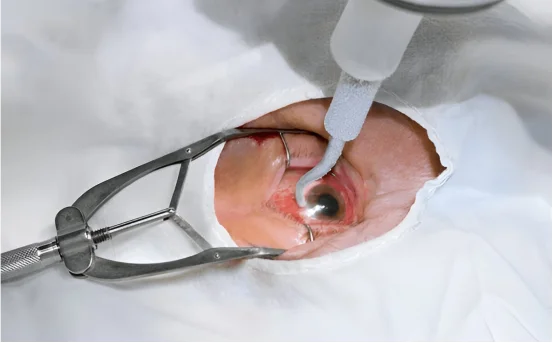
- Surgical Clipping
- The neurosurgeon places a metal clip at the base of the aneurysm.
- Prevents further bleeding permanently.
- Endovascular Coiling
- A catheter is inserted through the groin or wrist up to the brain.
- Coils are placed inside the aneurysm to block blood flow.
- A catheter is inserted through the groin or wrist up to the brain.
Factors that guide treatment choice
- Aneurysm size and shape
- Location in the brain
- Patient’s age and overall health
- Stability of the patient
- Presence of complications like hydrocephalus
In some cases, additional procedures such as external ventricular drainage (EVD) may be required to relieve pressure inside the brain.
Additional Tests After Initial Diagnosis
These tests help monitor recovery and detect complications
- Transcranial Doppler (TCD) evaluates vasospasm
- Blood tests check clotting factors, electrolytes, and infection markers
- Repeat CT scans monitor worsening or improvement
- ECG and cardiac enzymes because SAH can affect the heart
Monitoring continues for several days or weeks depending on severity.
Why Early Diagnosis Matters
Subarachnoid hemorrhage can worsen rapidly. Quick diagnosis ensures
- Reduced risk of rebleeding
- Early surgical intervention
- Better long-term neurological outcomes
- Prevention of stroke or brain swelling
- Higher survival rates
Delays in diagnosis can significantly increase complications and mortality.
Conclusion
The diagnosis of subarachnoid hemorrhage involves a combination of clinical evaluation, CT scans, angiographic studies, lumbar puncture, and advanced imaging techniques. Each test plays a vital role in confirming bleeding, identifying its cause, and planning life-saving treatments such as surgical clipping or endovascular coiling. Early detection is essential to prevent complications and improve outcomes.