Diagnosis of temporal lobectomy is a specialized brain surgery used to treat drug-resistant epilepsy that originates in the temporal lobe. It is one of the most common and effective surgical treatments for focal epilepsy. However, not every patient with epilepsy is an ideal candidate for this procedure. The decision to undergo a temporal lobectomy is only made after a comprehensive diagnostic evaluation to determine the exact location of seizure onset and assess whether surgery is likely to improve seizure control without causing major neurological deficits.
But undergoing brain surgery is a serious decision, and it isn’t made lightly. Before a patient is recommended for temporal lobectomy, a thorough and highly specialized diagnostic process must be conducted. This process is not only designed to confirm that the seizures originate in the temporal lobe but also to ensure that removing a portion of the brain will not impair critical functions like language, memory, or motor skills.
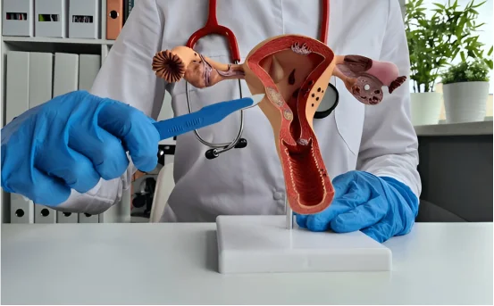
What Is Temporal Lobectomy?
Temporal lobectomy is a neurosurgical procedure that involves removing a portion of the temporal lobe typically the medial temporal structures, such as the hippocampus and amygdala to control seizures. This surgery is often recommended for individuals with medically refractory temporal lobe epilepsy (TLE), especially when seizures do not respond to two or more anti-seizure medications.
Temporal lobe epilepsy is the most common type of focal epilepsy, and when the seizures originate from a specific, localized area of the brain, surgery can offer substantial improvement—sometimes even a cure.
Why Is a Diagnosis Needed Before Temporal Lobectomy?
Brain surgery is a major step, and the risks must be carefully balanced against the potential benefits. To ensure the correct identification of the seizure focus and minimize the chances of complications, patients undergo a comprehensive diagnostic evaluation. The goal is to determine :-
-
The exact origin of the seizures
-
Whether seizures are localized to one side of the brain
-
Whether the brain region involved can be safely removed
-
The potential cognitive and functional effects of surgery
Only after these factors are evaluated can a neurologist and epilepsy surgery team recommend temporal lobectomy with confidence.
Diagnosis of Temporal Lobectomy
Here’s a step-by-step breakdown of the tests and evaluations typically performed :-
Detailed Clinical History and Seizure Description
The diagnostic journey begins with an in-depth review of the patient’s seizure history, including:
-
Age of seizure onset
-
Seizure frequency and duration
-
Triggers and patterns
-
Previous treatments and medications
Doctors will also examine the semiology of seizures how the seizures look and feel since certain seizure behaviors can suggest a temporal lobe origin.
Video Electroencephalogram (Video EEG) Monitoring
Video EEG is a cornerstone in epilepsy surgery evaluation. It involves :-
-
Continuous EEG recording (brain wave activity)
-
Simultaneous video monitoring to observe physical symptoms
Patients are admitted to an epilepsy monitoring unit (EMU) for several days while medications may be tapered to provoke seizures. This helps determine :-
-
The exact location of seizure onset (temporal lobe or elsewhere)
-
Whether the seizures are coming from one side or both sides of the brain
Clear, localized seizures from a single temporal lobe make a patient more likely to be a surgical candidate.
Magnetic Resonance Imaging (MRI) of the Brain
High-resolution MRI scans are performed to look for structural brain abnormalities, such as :-
-
Mesial temporal sclerosis (scarring of the hippocampus)
-
Brain tumors or cysts
-
Malformations of cortical development
MRI can also be enhanced with epilepsy protocols for more detailed imaging. A visible lesion in the temporal lobe that matches the seizure focus is a strong indicator for successful surgery.
Neuropsychological Testing
Cognitive testing assesses memory, language, attention, and problem-solving skills. These tests help :-
-
Localize brain function (e.g., which hemisphere is dominant for language)
-
Predict potential cognitive outcomes after surgery
-
Identify any baseline impairments that might worsen with surgery
For instance, in left temporal lobectomy candidates (typically the dominant hemisphere), language function is assessed thoroughly to avoid post-operative language deficits.
Functional MRI (fMRI) and Wada Test
These specialized tests evaluate language and memory dominance in the brain.
-
fMRI maps areas involved in language and memory using non-invasive imaging.
-
The Wada test involves injecting medication into one side of the brain to temporarily shut it down, assessing function on the other side.
This information helps determine whether removing the affected temporal lobe would compromise essential brain functions.
Positron Emission Tomography (PET) Scan
In patients whose MRI does not show obvious abnormalities, a PET scan can identify areas of decreased glucose metabolism in the brain, which may correspond to the seizure focus.
A temporal lobe with reduced activity on PET supports the diagnosis of temporal lobe epilepsy, especially when aligned with EEG findings.
Single Photon Emission Computed Tomography (SPECT)
SPECT imaging measures blood flow in the brain during or after a seizure. A type called ictal SPECT is done during a seizure and can highlight areas of increased blood flow that correspond to the seizure origin.
When combined with MRI in a technique known as SISCOM (Subtraction Ictal SPECT Coregistered to MRI), it offers a powerful way to visualize seizure focus in difficult-to-localize cases.
Psychiatric Evaluation
A mental health assessment is often performed to :-
-
Screen for mood disorders, anxiety, or depression
-
Ensure the patient has realistic expectations about surgery
-
Evaluate coping skills and psychosocial support
Emotional well-being plays an important role in post-surgical recovery and seizure outcomes.
Multidisciplinary Team Discussion
All findings from the diagnostic tests are reviewed by a comprehensive epilepsy surgery team, typically including :-
-
Epileptologists
-
Neurosurgeons
-
Neuropsychologists
-
Radiologists
-
EEG technologists
-
Social workers or psychiatrists
Together, they determine whether the patient is a good candidate for temporal lobectomy and if the benefits outweigh the potential risks.
When Is Temporal Lobectomy Recommended?
Temporal lobectomy may be recommended if :-
-
Seizures are focal and arise from the temporal lobe
-
Seizures are medication-resistant
-
Imaging and EEG clearly localize seizure onset
-
The epileptogenic zone is in a non-eloquent (non-critical) area of the brain
-
The patient is physically and mentally fit for surgery
Studies show that up to 70–80% of carefully selected patients become seizure-free after temporal lobectomy.
Conclusion
The diagnosis of temporal lobectomy candidacy involves more than just identifying seizures it’s about precisely localizing them, understanding brain function, and evaluating the impact of surgery on a person’s quality of life. This detailed diagnostic process ensures that the right patients are selected, reducing risks and maximizing the potential for seizure freedom.