Introduction
Vascular malformations are abnormal formations of blood vessels that are present at birth, though they may not be visible immediately. Unlike vascular tumors, such as hemangiomas, vascular malformations grow proportionally with the individual and do not regress over time. Early detection and accurate diagnosis are critical for effective management and treatment. These conditions can affect veins, arteries, capillaries, or lymphatic vessels, and their symptoms may range from mild cosmetic concerns to life-threatening complications. Understanding how vascular malformations are diagnosed helps patients access timely treatment and improves outcomes.
What Are Vascular Malformations?
Vascular malformations are structural anomalies of the blood vessels that occur during fetal development. They are typically classified based on the type of vessels involved: capillary, venous, arterial, lymphatic, or combined types. Unlike vascular tumors, malformations do not show rapid growth in infancy but persist throughout life. They can appear as swelling, discoloration, or deformity in the affected area. While some vascular malformations are minor and only cosmetic, others may cause pain, bleeding, or organ dysfunction, depending on their location and size.
Importance of Accurate Diagnosis
Diagnosing vascular malformations accurately is essential for several reasons:
- Tailored Treatment: Correct identification ensures that the treatment plan targets the specific type of malformation.
- Avoiding Misdiagnosis: Some vascular malformations resemble other conditions such as cysts, hematomas, or tumors. Misdiagnosis can lead to inappropriate treatments.
- Monitoring Complications: Proper diagnosis helps monitor potential complications, including thrombosis, ulceration, or hemorrhage.
- Surgical Planning: Knowing the exact type, size, and flow characteristics of the malformation is crucial for planning any surgical or interventional procedure.
Clinical Examination
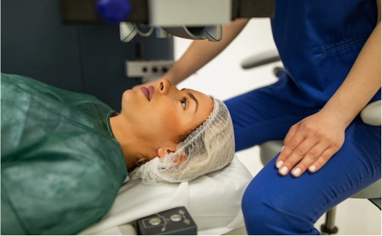
The diagnosis of vascular malformations begins with a thorough clinical evaluation by a specialist, usually a vascular surgeon, interventional radiologist, or dermatologist. The clinical examination includes:
- Patient History: Doctors inquire about the lesion’s onset, growth patterns, associated symptoms like pain or bleeding, and family history of similar conditions.
- Physical Examination: The affected area is examined for size, texture, color, warmth, tenderness, and pulsatility. Low-flow malformations like venous or lymphatic types often feel soft and compressible, while high-flow malformations like arteriovenous malformations feel firm and may exhibit a thrill or bruit.
- Functional Assessment: If the malformation affects limbs or vital organs, doctors assess movement, strength, and functionality to determine the severity of impact.
Imaging Techniques
Imaging plays a central role in diagnosing vascular malformations. Various modalities help determine the type, extent, and blood flow characteristics of the malformation:
- Ultrasound with Doppler :- Ultrasound is often the first imaging test used. Doppler ultrasound helps visualize blood flow within the lesion, distinguishing high-flow malformations (arteriovenous) from low-flow types (venous or lymphatic). This technique is non-invasive, quick, and widely available.
- Magnetic Resonance Imaging (MRI) :-MRI provides detailed images of soft tissues and is highly effective in mapping the extent of the malformation. MR angiography (MRA) allows visualization of arteries and veins, aiding in the assessment of complex or deep lesions. MRI is especially useful for planning surgical or interventional treatments.
- Computed Tomography (CT) Scan :- CT scans, including CT angiography, are helpful in evaluating bone involvement or complex vascular anatomy. Although less preferred than MRI for soft tissue analysis, CT is valuable in certain anatomical regions or for rapid assessment in emergencies.
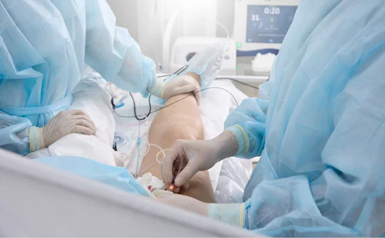
- Conventional Angiography :- This invasive technique involves injecting contrast dye into the blood vessels to visualize the precise vascular architecture. Angiography is often used when an interventional procedure, like embolization, is being considered. It helps map feeding arteries, draining veins, and the malformation’s flow dynamics.
- Lymphangiography :- For lymphatic malformations, specialized imaging like lymphangiography or MRI lymphangiography is employed. These techniques help identify cystic spaces and connections to lymphatic channels.
Laboratory Tests
Although imaging is the mainstay for diagnosis, certain laboratory tests may be indicated in specific cases:
- Coagulation Profile: Venous malformations can be associated with localized intravascular coagulation, necessitating evaluation of clotting factors.
- Complete Blood Count (CBC): To check for anemia or infection if there is bleeding or ulceration.
- Genetic Testing: In rare cases, genetic tests may be advised to identify syndromes associated with vascular malformations, such as CLOVES or Klippel-Trénaunay syndrome.
Classification and Diagnostic Criteria
Once all evaluations are complete, vascular malformations are classified based on flow characteristics and involved vessels:
- Low-Flow Malformations: Includes venous, capillary, lymphatic, or combined types. These are generally slow-growing and less symptomatic.
- High-Flow Malformations: Includes arterial or arteriovenous malformations, which are prone to rapid growth, bleeding, and cardiovascular complications.
Accurate classification guides treatment decisions. For example, sclerotherapy is often effective for low-flow venous malformations, while high-flow arteriovenous malformations may require embolization followed by surgical intervention.
Challenges in Diagnosis
Diagnosing vascular malformations can be challenging due to:
- Similar Appearance: Some lesions resemble benign cysts, tumors, or hematomas.
- Deep Lesions: Malformations located deep in muscles or organs may not be visible externally, requiring advanced imaging.
- Variable Symptoms: Symptoms may develop slowly, leading to delayed diagnosis.
- Complex Anatomy: Mixed malformations involve multiple vessel types, complicating assessment and treatment planning.
Multidisciplinary Approach
Given the complexity of vascular malformations, a multidisciplinary approach is often required. Teams typically include:
- Vascular surgeons
- Interventional radiologists
- Dermatologists
- Plastic and reconstructive surgeons
- Genetic counselors
Collaboration ensures comprehensive evaluation, precise diagnosis, and personalized treatment planning.
Conclusion
Early and accurate diagnosis of vascular malformations is vital to prevent complications, improve outcomes, and enhance quality of life. A combination of thorough clinical examination, advanced imaging, and laboratory tests allows specialists to classify malformations and tailor treatment strategies effectively. Awareness and timely consultation with vascular specialists are key steps for anyone affected by these congenital vascular anomalies.