Enucleation is the surgical removal of the entire eyeball (globe) and its contents. This eye removal surgery is done only when an eye is irreversibly damaged or diseased. Common reasons include severe eye cancer or trauma that cannot be treated any other way. After the eyeball is taken out, an artificial prosthetic eye is usually fitted to restore a natural appearance. In practice, enucleation eliminates a non-functioning, painful, or dangerous eye to protect a patient’s overall health or relieve suffering.
Why is Enucleation Surgery Performed?
Enucleation is considered a last resort to save health or relieve pain when the eye cannot be saved by any other treatment. Doctors recommend it in cases where the eye is causing more harm than good. For example, the main reasons for eye removal include uncontrolled pain and preventing the spread of disease In practice, that means:
- Eye cancer or tumors: Dangerous intraocular tumors (such as melanoma or childhood retinoblastoma) often require removal of the eye to prevent cancer from spreading.
- Severe trauma: A major injury that shatters the eyeball can leave it beyond repair. Enucleation prevents long-term pain or infection in an eye that no longer works.
- Uncontrolled infection: An infection inside the eye (like endophthalmitis) that is not cured by medication may only be stopped by removing the infected tissue.
- Blind, painful eye: A non-seeing eye that causes constant discomfort or is disfigured is often removed to improve quality of life. For example, Cleveland Clinic notes that one main reason for enucleation is to relieve pain from an irreversibly diseased eye when other treatments have failed.
In the United States, the top cause of eye enucleation is trauma (eye injuries), followed by eye cancers. (In very young children, retinoblastoma is the most common reason.) In each case, surgeons carefully weigh the decision, since enucleation is only done when absolutely necessary.
Symptoms of Enucleation Surgery
“Symptoms of enucleation surgery” refers to warning signs that an eye problem is serious enough to need removal. These symptoms are actually signs of the underlying condition, and they include:
Intense, chronic eye pain: Especially in an eye that already has no vision. For instance, a “blind painful eye” that hurts constantly is a classic indication for removal.
Complete vision loss: Sudden or total loss of vision in one eye with no chance of recovery.
Persistent infection or inflammation: Ongoing redness, swelling, or discharge in the eye socket that does not clear with treatment.
Visible tumor or growth: A dark tumor in the eye, or anything protruding/abnormal on the eye.
Severe disfigurement: An eye that is visibly collapsed (phthisis bulbi), bulging, or cosmetically deforming.If a patient has any of these symptoms, an eye specialist will investigate immediately. These warning signs suggest the eye is beyond repair and may be causing systemic issues (pain, risk to the other eye, or spreading disease) that justify enucleation.
Causes of Enucleation Surgery
The causes of enucleation are the specific conditions that damage the eye so severely that it must be removed. They overlap with the reasons above but can be listed as:
- Eye trauma: Accidents (blunt force, cuts, burns) that irreparably injure the eye.
- Intraocular cancer: Malignancies inside the eye (e.g. melanoma, retinoblastoma) that threaten vision or life.
- Severe infection: Bacterial or parasitic infections (such as endophthalmitis or acanthamoeba keratitis) that destroy eye tissues.
- End-stage disease: Phthisis bulbi (a shrunken, scarred eye) often from long-term injury or disease, or congenital anomalies like microphthalmia (a very small eye).
- Autoimmune attack: Sympathetic ophthalmia, a rare condition where an injury in one eye triggers inflammation in both eyes, can force removal of the injured eye to protect the other.
In short, any condition that leaves the eye blind, painful, and untreatable – or that poses a danger of spreading (cancer/infection) – can cause doctors to recommend enucleation.
Diagnosis of Enucleation Surgery
Before deciding on enucleation, doctors perform a thorough diagnosis. This typically includes:
- Comprehensive eye exam: The ophthalmologist checks vision (confirming the eye has no useful sight) and examines all internal and external eye structures.
- Imaging tests: Ultrasound, CT or MRI scans may be done to evaluate the extent of a tumor, the degree of internal damage, or hidden complications.
- Laboratory tests: If infection is suspected, samples from the eye may be cultured or other lab tests may be done. If cancer is suspected, a biopsy or specialized imaging helps confirm the diagnosis.
- Review of alternatives: Doctors will try all possible treatments first (like laser therapy for retinoblastoma or intensive antibiotics for infection). They confirm that saving the eye is not feasible.
In many cases (such as large retinoblastoma tumors), doctors only proceed to enucleation if there is essentially no chance of useful vision left in the eye. If any vision can be saved, less drastic treatments are attempted first. Once it is clear that the eye is beyond help, the medical team will carefully plan the enucleation surgery, including discussing the steps and risks with the patient.
Treatment of Enucleation Surgery
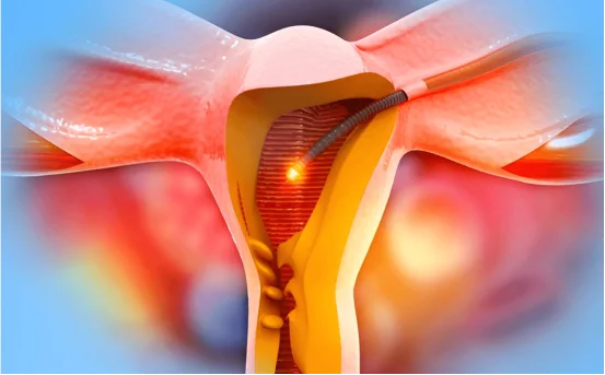
The Eye Enucleation Procedure: Enucleation is performed in an operating room under anesthesia. The surgery usually takes about 1–2 hours. Here’s what happens during the procedure:
- The surgeon makes an incision in the eyelids and carefully separates the eye muscles. They detach the four extraocular muscles and cut the optic nerve to free the eyeball.
- The entire eyeball (globe) is removed from the orbit.
- A spherical orbital implant (often silicone or porous material) is immediately placed in the empty eye socket to fill the space and maintain normal shape.
- The remaining eye muscles are stitched to the implant. This allows the eventual prosthetic eye to move more naturally.
- A conformer (a smooth plastic shell) and/or a pressure bandage is placed over the implant to keep everything in the proper position as the socket heals. The surgeon may temporarily stitch the eyelids partially closed to protect the area.
After surgery, the patient is taken to recovery. Most people go home the same day or within 24 hours. Pain is usually mild; over-the-counter pain relievers (like acetaminophen or ibuprofen) are often sufficient, and any discomfort typically lasts only a few days. Patients are given detailed care instructions, including how to keep the area clean and how to use any prescribed antibiotic drops. Strenuous activity, heavy lifting, or bending over is generally avoided for several weeks to ensure proper healing.
Approximately one week after surgery, the bandage and conformer are removed for examination. About 6–8 weeks later, once healing is complete, the patient will work with an ocularist (an eye prosthesis specialist). The ocularist takes an impression of the healed socket and creates a custom artificial eye that matches the other eye in size and color. When inserted, this prosthetic eye sits over the implant and under the eyelids, giving a natural appearance.
Enucleation vs Evisceration
Sometimes eye removal can be done by evisceration instead of enucleation. The differences are:
- Enucleation: Removes the entire eyeball (globe) from the socket, including the white sclera and all internal contents. The extraocular muscles and optic nerve are cut, then later reattached to the implant.
- Evisceration: Removes only the inner contents of the eye (cornea, iris, retina, lens), leaving the white sclera and outer wall intact. It is less extensive and often faster.
Evisceration is only an option when the eye disease is limited (for example, it is never done if a cancer is present, because diseased tissue must be completely removed). In general, enucleation is preferred if there is a tumor or risk of spreading infection. It also best prevents sympathetic ophthalmia (a rare immune condition that can attack the healthy eye after serious injury). As Michigan Medicine explains, evisceration “is a less invasive procedure and may be an option depending on the condition of the eye,” but the surgeon will advise which approach is safest.
Conclusion
Enucleation surgery is a major procedure, but it can be life-changing for the better when an eye is beyond repair. It effectively removes sources of pain, infection, or cancer, even though it means losing vision in that eye. Thanks to modern techniques (like orbital implants and well-crafted prosthetic eyes), most patients heal well and regain a normal appearance. Importantly, by removing a diseased eye, enucleation often provides significant pain relief and treats or prevents serious disease.
If you or a loved one face this surgery, work closely with your medical team. They will explain each step — from the eye removal procedure itself to recovery and prosthetic fitting — so you know what to expect. With proper care, follow-up, and support, many patients adapt smoothly and return to daily activities in a few weeks.