In the last few time Neurosurgery seen a major transformation providing patients with better, quicker and more effective treatments for spinal and brain-related diseases. A major and groundbreaking innovations in this area is Neuroendoscopy Surgery, a minimally invasive surgical procedure that permits neurosurgeons to reach and treat specific areas of the brain via small openings made by an endoscope.
Neuroendoscopy uses an extremely thin and flexible tube with a camera and a light — referred to as an endoscope to navigate inside the brain. It gives real-time video and permits the surgeon to perform surgery with extreme accuracy and reduce the amount of trauma that is typically experienced during traditional open-brain surgery. This cutting-edge technique is typically employed to treat ailments like hydrocephalus, the brain, tumors of colloid or pituitary gland tumours.
Why Neuroendoscopy Surgery?
There are many compelling reasons for why neuroendoscopy is preferable to traditional brain surgery in a variety of cases:
- Minimally Invasive:- Contrary to traditional surgeries that require the use of a huge skull surgery called craniotomy (opening of the skull) Neuroendoscopy is a procedure that requires just an incision of a tiny size. This means less discomfort, a lesser bleeding, and a lower chance of developing an infection.
- Faster Recovery:- Patients who undergo neuroendoscopic procedures typically will have lower hospital stays and quicker recovery times, and quicker return to routine.
- Lower Complication Rates:- The improved visibility provided by the endoscope minimizes the chance of causing damage to brain tissue. This means lower rates of complications and better surgical results.
- Cosmetic Advantage:- Incisions that are smaller mean less scarring which is an important factor for a lot of patients.
- Precision in Complex Areas:- Neuroendoscopy can be extremely beneficial in accessing complex or deep brain areas that would otherwise be difficult or dangerous to access via open surgery.
Symptoms Indicating the Need for Neuroendoscopy Surgery
While not every neurological sign necessitates surgery, certain signs might indicate the need for neuroendoscopic surgery. The symptoms vary based on the cause, but may include:
- Chronic migraines (often because of pressure building-up inside the brain)
- Nausea and vomiting
- Double vision or blurred
- Balance or coordination issues
- Seizures
- Memory problems
- Cognitive or behavioral changes
- Signs of hydrocephalus, such as an the head growing larger in infants, or difficulties walking in adults
These symptoms could be indicative of problems like brain cysts, tumors or blockages in cerebrospinal fluid (CSF)–all of which can be efficiently treated by neuroendoscopy.
Causes Leading to Neuroendoscopy Surgery
The root causes for needing neuroendoscopy surgeries usually involve disorders in brain function or structure that are not treatable medically. The most common causes are:
- Hydrocephalus: A deficiency in the accumulation of CSF in the brain. usually treated by an endoscopic 3rd ventriculostomy (ETV).
- Brain tumors: Particularly colloid cysts, ependymomas or pituitary tumors, which are found in the midline or deep areas of the brain.
- The tumors of the intraventricular space: inside the ventricles of the brain.
- Cysts: These include colloid or arachnoid cysts that can block CSF flow.
- Clot or hemorrhage In certain instances an endoscopy that is minimally invasive can aid in removing hemorrhage-related blood clots.
Intervention and early detection are essential to effectively managing these conditions and avoiding any complications.
Diagnosis Before Neuroendoscopy Surgery
Before deciding to undergo neuroendoscopy, it is necessary to conduct a thorough diagnosis performed to determine the type and extent of the issue. The most common diagnostic steps are:
- Neurological Examination:- Initial tests test the cognitive function, reflexes, visual abilities, motor skill and coordination, to determine symptoms of brain dysfunction.
- Imaging Tests:- The most advanced imaging technologies are utilized to identify the problem:
- MRI (Magnetic Resonance Imaging) Provides clear views of brain tissue and can help to identify tumors or blockages.
- CT Scan (Computed Tomography): Useful for emergencies, particularly in detecting swelling, bleeding or bruising.
- Endoscopic Examination: in certain instances, exploratory endoscopy could be used to examine the region prior to surgery.
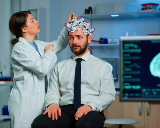
- Electroencephalogram (EEG):- If seizures are part the symptoms pattern, EEG helps track brain activity.
- Lumbar Puncture (Spinal Tap):- To study cerebrospinal fluid under conditions such as hydrocephalus or infections.These diagnostic techniques assist the surgeon to determine if neuroendoscopy is the right procedure.
Treatment Through Neuroendoscopy Surgery
If a problem is identified and surgery is recommended Neuroendoscopy can be a safer and more effective treatment option. The procedure generally is carried out:
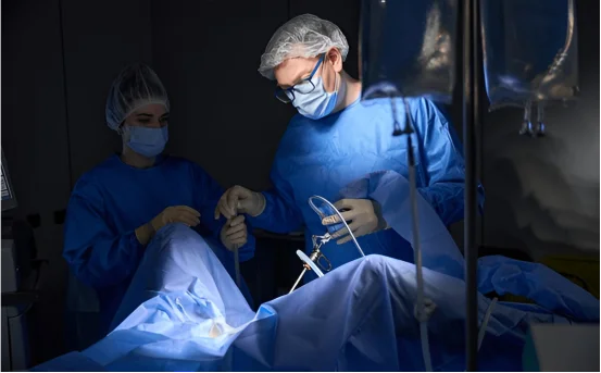
Procedure Overview:
- Performed under general anesthesia.
- A tiny opening (burr hole) is made in the skull.
- An endoscope is used to view the targeted area.
- Specialized surgical instruments are utilized via the endoscope to eliminate tumors and to create pathways to CSF or biopsy tissues.
The endoscope is then removed and the cut is closed with a minimal suturing.
Types of Neuroendoscopic Procedures:
Endoscopic 3rd Ventriculostomy (ETV) typically employed to treat hydrocephalus involves forming a small hole inside the third ventricle’s floor to clear the CSF obstruction.
- Tumor Resection Removal of pituitary or intraventricular tumors by using an endoscope.
- Cyst Fenestration: The process of creating an opening in the cyst wall that allows the drainage of fluid.
- Biopsy: Collection of tissue samples to analyze when the tumor’s type is not known.
Post-Surgical Care:
- Patients usually stay in the hospital for between 1-3 days.
- There is no discomfort or pain because of the less invasive nature of the procedure.
- Neurological evaluations and follow-up scans to monitor the recovery.
In some instances patients might require physiotherapy or prescription medications to control seizures or reduction in swelling.
Conclusion
Neuroendoscopy Surgery represents a paradigm change in the treatment for difficult brain conditions. It is a combination of cutting-edge technology and the expertise of a surgeon to offer an effective, minimally-invasive treatment that significantly improves outcomes for patients. With less recovery time as well as fewer risks and high precision, this method is now the standard for a variety of neurosurgical conditions.
If you or someone close to you is suffering from a health issue, speak with an experienced neurosurgeon to determine whether neuroendoscopy is the best option. The early detection, the use of modern imaging and minimally-invasive interventions can make a huge difference in ensuring your patient’s safety and effective recovery.