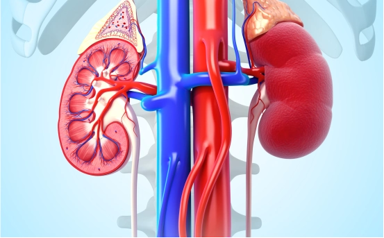
Ureteroscopy is a minimally invasive procedure used to diagnose and treat conditions affecting the ureters the thin tubes that carry urine from the kidneys to the bladder. This technique has become a standard approach in urology, especially for detecting and removing kidney stones and diagnosing abnormalities in the urinary tract.
Ureteroscopy has become a widely preferred technique in modern urology due to its precision, safety, and rapid recovery times. Using a thin, tube like instrument called a ureteroscope, doctors can directly visualize the inside of the ureter and kidneys to identify and remove stones, biopsy abnormal tissues, or relieve obstructions all without making a single incision on the body.
What is Ureteroscopy?
Ureteroscopy is a procedure where a thin, flexible or rigid instrument called a ureteroscope is inserted into the urethra and passed through the bladder into the ureter. The ureteroscope allows the urologist to visually examine the inner walls of the ureter and the kidney. It is commonly used for diagnosing and treating ureteral stones, tumors, strictures, or unexplained bleeding.
Unlike open surgeries, ureteroscopy does not require incisions, making it less invasive and associated with a quicker recovery time.
Why is Ureteroscopy Performed?
The most common reason for performing ureteroscopy is to remove ureteral or kidney stones that are too large to pass on their own. In addition, this procedure is used for :-
-
Investigating hematuria (blood in urine)
-
Diagnosing tumors or polyps in the urinary tract
-
Evaluating abnormalities detected through imaging tests
-
Removing or biopsying abnormal tissue
-
Treating strictures (narrowing of the ureter)
Types of Ureteroscopes Used
There are two primary types of ureteroscopes :-
-
Rigid Ureteroscope :- Used for examining and treating the lower ureter near the bladder. It offers excellent image clarity and better control during stone removal.
-
Flexible Ureteroscope :- Designed to reach higher into the ureter and kidneys. It can bend and flex to access difficult to reach areas and is ideal for navigating through tight curves.
The choice of instrument depends on the location and size of the stone or abnormality, as well as patient-specific factors.
Preparing for the Procedure
Before undergoing ureteroscopy, patients will typically have a consultation with a urologist who may recommend blood tests, urine tests, and imaging studies like ultrasound, CT scan, or X-ray to determine the exact location and size of the stones or abnormalities.
Patients are advised to :-
-
Avoid eating or drinking for several hours before the procedure
-
Stop certain medications that could increase bleeding risk (as directed by the doctor)
-
Arrange transportation, as sedation or anesthesia is usually required
A complete medical history will also be reviewed to minimize any potential complications.
Procedure of Ureteroscopy
- Anesthesia Administration :- Ureteroscopy is usually performed under general or spinal anesthesia, ensuring that the patient feels no discomfort during the procedure. In some cases, local anesthesia with sedation may also be used.
- Insertion of the Ureteroscope :- Once the anesthesia takes effect, the urologist gently inserts the ureteroscope through the urethra and passes it into the bladder. From there, it is directed into the ureter and sometimes into the kidney, depending on the location of the issue.
- Visual Inspection and Treatment :- The ureteroscope transmits live images to a monitor, allowing the surgeon to inspect the urinary tract in detail. If stones are detected, they may be fragmented using a laser (typically Holmium laser) and the pieces are removed using a basket like tool. In case of tumors or suspicious tissue, a biopsy may be taken for further analysis.
- Stent Placement (If Needed) :- In certain cases, a temporary stent (a thin tube) may be placed in the ureter to ensure proper urine flow and reduce swelling or discomfort. The stent is typically removed after a few days or weeks.
- Completion and Recovery :- After completing the inspection or treatment, the ureteroscope is removed. The entire procedure usually lasts between 30 minutes to an hour, depending on the complexity.
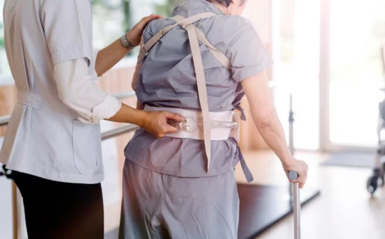
Recovery After Ureteroscopy
Most patients can go home the same day after the procedure. However, some may be observed in the hospital for a few hours, especially if a stent was placed or if complications are anticipated.
Post operative instructions include :-
-
Drinking plenty of water to help flush out the urinary tract
-
Taking prescribed pain relievers or antibiotics
-
Avoiding heavy lifting or strenuous activity for a few days
-
Noticing mild blood in the urine, which is normal for 24–48 hours
If a stent is placed, mild discomfort or a frequent urge to urinate may occur until the stent is removed.
Risks and Complications
Although ureteroscopy is considered safe, some risks may include :-
-
Urinary tract infection (UTI)
-
Temporary discomfort during urination
-
Bleeding or injury to the ureter
-
Stent-related irritation
-
Rarely, ureteral perforation or stricture formation
These complications are uncommon and can usually be managed with prompt medical attention.
Benefits of Ureteroscopy
Ureteroscopy offers several advantages over traditional surgical methods :-
-
No external cuts or stitches
-
Short recovery time
-
High success rate for stone removal
-
Ability to treat both ureteral and kidney conditions
-
Suitable for patients who are not candidates for other treatments like shockwave lithotripsy
Because it is minimally invasive, most patients return to normal activities within a few days.
Conclusion
Ureteroscopy is a highly effective and minimally invasive procedure for diagnosing and treating conditions within the urinary tract, especially kidney and ureteral stones. With advancements in endoscopic technology, it has become a preferred treatment method due to its accuracy, safety, and fast recovery.