The issue of retinal detachments is becoming more common in society. Visually, it is an issue that requires a lot of medical attention. Retinal detachment occurs when the delicate tissues of the eye begin to separate from the eyeball. If left untreated, it may result in irreversible blindness. One of the possible surgical treatments in these symptoms for retinal detachment surgery.
What is Retinal Detachment?
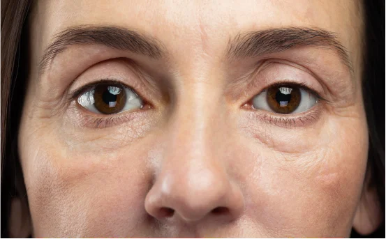
Understanding of the symptoms for retinal detachment surgery
Before we proceed with problems, it would be crucial to discuss some eye anatomy basics and the consequent layers of a retina. Detaching or sustaining an injury is something people would want to avoid, but if prevention measures are not taken, they risk partial or complete vision loss.
There are individuals who seek to wear aesthetic contact lenses but end up developing certain eye problems which necessitates surgical intervention to restore normalcy.
Types of Retinal Detachment
There are three major types of retinal detachment which include:
- Rhegmatogenous Retinal Detachment:- This is the most common form and it happens when a fluid filled space forms underneath the retina that is separated by some portion and is caused by a tear or break in the retina.
- Tractional Retinal Detachment:- This occurs when scar tissue is formed over the surface of the retina, as is the case with diabetic retinopathy.
- Exudative Retinal Detachment: –This occurs when fluid accumulates beneath the retina but there is no associated tear. Tumors, abnormal growths, inflammation, or even abnormal blood vessels can lead to this condition.
Common Causes of Retinal Detachment Surgery
1. Retinal Tears or Holes
Most common complaint of eye problems is a tear in the retina, where an eye pulling away results in a certain degree of separation. This very frequently happens as one ages, and a tear typically occurs as fluid starts to segregate. After a tear, the retina has to be surgically fixed.
2. Aging and Posterior Vitreous Detachment (PVD)
With increasing age, most people will undergo changes in the shape and size of their eyes as a result of a gel-like substance in the eye expanding an area within the eye. While this is beneficial for the retina since it can help in the adhesion of the retina to the vitreous posterior region, it can also turn the retina into a structure that is more easily breakable if there are strong attachments of vitreous to it.
Individuals over 50 years of age are more susceptible to having a PVD, thus increasing the likelihood of retinal tears and detachment.
3. Severe Myopia (Nearsightedness)
People suffering from high myopia have an elongation of the eyeballs which leads towards the stretching and thinning of the retina. This makes it much easier to tear, and to develop holes or detachment. Moreover, those undergoing higher degrees of myopia tend to undergo cataract surgery more frequently.
4. Eye Trauma or Injury
Both blunt and penetrating eye injuries can result in the separation of the retina. These can be sustained through participation in sports or through motor vehicle accidents and work-related injuries. In these situations, timely emergency surgery for retinal detachment is critical for preventing irreversible vision loss.
5. Previous Eye Surgery
Cataract or laser eye surgical procedures place a person at slightly higher risk for retinal detachment. The postoperative inflammation or the surgical techniques may cause vapor bubbles to form within the eye that could ultimately lead to detachment.
6. Diabetic Retinopathy
Diabetes that is not well managed can lead to scar tissue formation on the retina (proliferative diabetic retinopathy). This type of scarring may create a pulling force on the retina causing tractional retinal detachment. Surgery to remove the scar tissue and reattach the retina is performed in these cases.
7. Family History
Having a family member who is affected by retinal detachment increases the risk factors. Close relatives with surgical myopia also increase the chances of undergoing eye surgery.
8. Lattice Degeneration
Lattice degeneration is the thinning of the peripheral retina. It occurs in 10% of the population but may pose a risk by predisposing the retina to tears that lead to detachment.
9. Inflammatory Eye Diseases
Some types of uveitis and scleritis are known to cause the separation of layers beneath the retina, leading to exudative detachment. Chronic inflammation can weaken the adherence of the retina to the layers beneath it. While this can be fixated with surgery, the separation itself can be repaired only surgically.
Surgical Symptoms
Signs of retinal detachments are preventable if acted upon in a timely manner. Their detection reveals several discernible indicators:
- Unilateral or bilateral light emission in flashes
- Curtain-like obscurations of some portions of the visual field
- Acute appearance of floaters
- Vision distortion or blurriness
You should contact an ophthalmologist immediately if any of these concerns apply to you.
Diagnosing Retinal Detachment
Prompt action is crucial when addressing potential sight loss and requires the following measures:
- A dilated eye examination can identify torn or detached retinas.
- Ocular Ultrasound- Particularly useful when the retina is not directly visible.
- Optical Coherence Tomography (OCT)- High-resolution imaging designed to detect fluid accumulation or separation of the retina.
Post-diagnosis, the ophthalmologist will decide on the necessity and type of surgery to be conducted for retinal detachment.
What are the reasons for surgical intervention for retinal detachments?
Neglecting a retinal detachment can lead to irreversible vision loss for an individual. Surgical intervention helps to:
- Restore the anatomical position of the retina.
- Close the retinal tears and holes.
- Removal of scar tissue and accumulated fluids.
Some of the more commonly known approaches include:
- Pneumatic Retinopexy
- Scleral Buckling
- Vitrectomy
Treatment varies according to the type and degree of detachment as well as the ocular health of the patient.
Conclusion
Timely surgical intervention is paramount for retinal detachment, a condition that endangers vision. Anatomy and physiology of the eye is ne cessary to understand for effective early detection and prevention. Risk factors include a history of eye trauma, severe myopia, age-related changes, diabetic retinopathy, and older age.
Individuals with sudden vision decline, new onset floaters, or flashes of light are cautioned not to ignore these changes. Preservation of vision is possible with early diagnosis and prompt surgery.