Your eyes are the portals through which you experience the world, yet serious internal problems can jeopardize that view. One such threat is symptoms for retinal surgery or other disorders that sometimes make surgery necessary-a crucial operation designed to save sight. Recognizing the symptoms for retinal surgery early and knowing when an operation is unavoidable can mean the difference between healing fully and losing vision for good. In the paragraphs that follow, we explain the red flags, their importance, and the actions you should take.
The retina is a thin, light-sensitive layer that lines the inside back of the eye. It converts incoming light into nerve signals and sends those signals to the brain, where images are created. If the retina pulls away from its underlying tissue-a situation called retinal detachment-damage can spread quickly, sometimes resulting in permanent blindness.
- Detached areas no longer receive oxygen or nutrients, and those cells start to die.
- Because the macula is responsible for sharp central vision, its involvement greatly raises the stakes.
- When care is postponed, the odds of restoring full sight plummet, so speed matters.
Retina surgeons often say that even quick in-office fixes such as pneumatic retinopexy or laser treatment must be done without delay to stop progression. Knowing the early symptoms lets you act sooner, and that timeliness can greatly improve the results of any necessary surgery.
Being aware of the symptoms for retinal surgery is essential for timely intervention. This knowledge can empower you to seek help before permanent damage occurs.
Core Symptoms That May Flag Retinal Problems
Sudden Surge of Floaters
- Floaters-small spots, threads, or spider-web wisps that drift through sight-often multiply as people age. Yet a sudden shower or swarm points to possible posterior vitreous separation or even a retinal tear.
- That change happens when the gel-like vitreous pulls free from the light-sensitive retina, risking further injury.
Bright Flashes of Light (Photopsias)
- Brief bursts or arcs of light-seen mostly in dim rooms-happen when the moving gel tugs at or tears the retinal surface.
- Though painless, these flashes demand prompt examination, since they may prelude more serious loss.
Curtain or Shadow Across Vision
- Perhaps the clearest warning of detachment, a dark curtain, wedge, or veil creeps across peripheral or central vision.
- It often begins at the edge and steadily broadens as healthy tissue peels away from the back of the eye.
Abrupt Blurring or Wavy Vision
- Fast blurring or distortion can signal that the macula or the area near the center of the retina is involved.
- Patients frequently describe wavy lines, faded colors, or sudden trouble reading, recognizing faces, or fixing on objects.
Loss of Peripheral (Side) Vision
- Loss of side vision is a serious warning sign. It may signal extensive retinal detachment and should be treated as an emergency.
Eye Discomfort, Headache, or Nausea
- Retinal detachment is usually painless, yet some people report a dull ache, often paired with a headache or mild nausea, particularly when tears develop or the detachment occurs quickly.
Washed-Out Colors, Poor Night Vision
- Though rarer, these changes may point toward diabetic or tractional detachment linked to diabetic retinopathy.
When to See a Specialist (and Why Surgery May Be Needed)
Immediate Red Flags
Consult a retina specialist or visit the E.R. without delay if you notice:
If you experience any of the symptoms for retinal surgery, do not hesitate to reach out to a healthcare provider to discuss your concerns.
- A sudden swarm of floaters
- New flashes or streaks of light
- A dark curtain or moving shadow obscuring part of your field
- A sharp increase in blur or loss of side vision.
Such signs imply that tearing or detachment could already be underway-and quick diagnosis and treatment are vital to protect your sight.
Understanding the symptoms for retinal surgery can help mitigate the risks associated with delayed treatment.
Diagnostic Steps
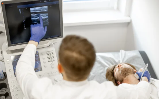
To confirm the problem, the ophthalmologist will perform a dilated exam and may use special lenses or ultrasound to find tears or fluid under the tissue.
Additional imaging-retinal OCT, fundus photos, or B-scan ultrasound-can provide further detail.
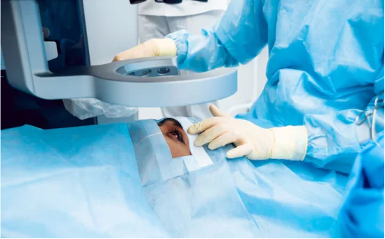
Surgical Treatments Explained
When a tear or detachment threatens vision, the exact procedure depends on how bad the damage is and where it occurs:
- *Laser photocoagulation or cryopexy (freezing): a quick blast of light or ice that seals tiny cracks before fluid spreads.
- *Pneumatic retinopexy: a small gas bubble is injected so it gently holds the tissue against the back of the eye. surgeons usually add a laser or freeze treatment to strengthen the patch.
- *Scleral buckling: a soft silicone band stitched around the eyeball pushes outward, easing the inward tug of a shrinking gel.
- *Vitrectomy: the thick gel is removed, trapped fluid is drained, and the cavity may be filled with gas or long-lasting oil.
Many of these office-based surgeries take an hour and require only numbing drops, yet complex cases still need a full OR and local or general anesthesia.
Timing Is Critical
Early action protects sight; every hour lost lets the tear widen and the macula slip. if the central zone is still attached at surgery, chances of full recovery climb sharply; once it lifts, the outcome turns hazy.
Overall success rates hover around ninety to ninety-five percent, and those odds improve the sooner patients arrive.
Conclusion
Awareness of symptoms for retinal surgery is crucial to preserving eye health and maintaining quality of life.
Retinal surgery is lifesaving work that tries to keep peoples vision intact when severe tears or detachments threaten permanent loss. noticing and reporting warning signs-sudden floaters, flashes, a curtain across the field, blurred objects, or fading edges-gives the best shot at complete restoration.
Swift diagnosis remains the highest priority. A careful dilated exam, paired with imaging tools such as OCT or ultrasound, lets eye doctors spot tears or detachments while they are still small. If intervention occurs before the macula lifts, procedures-from laser photocoagulation or cryopexy to pneumatic retinopexy, scleral buckling, or vitrectomy-practice show an anatomical success rate exceeding 90 percent and deliver noticeably sharper vision. Recognizing the symptoms for retinal surgery early can significantly impact treatment success and visual outcomes.
Nonetheless, diligent aftercare is equally vital. Simple habits such as keeping the head in the prescribed position, steering clear of heavy lifting, and rigorously timing the eye drops greatly boost outcomes and minimize trouble. Even when several surgeries become necessary, todays microsurgical methods often restore both the eye’s normal shape and useful sight.