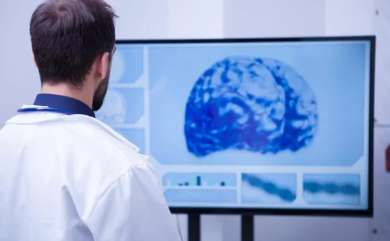
Retinal causes for retinal surgery, especially tears and detachments, threaten eyesight and must be diagnosed and treated without delay. The retina is a fragile, light-sensitive film at the rear of the eye that changes incoming light into nerve impulses for the brain. Damage from a tear, scar tissue, or fluid under the membrane can let the retina peel away from its bed, ruining vision and raising the risk of partial or total blindness.
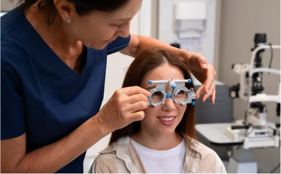
Warning signs may be quick: flashing lights, floaters, blurred patches, or a curtain gathering at the edge of sight. Noting them and getting professional help right away is crucial; these disorders deteriorate rapidly, and the good window for fixing the problem is often only hours long.
Why Treatment and Causes for Retinal Surgery Matters
- Prevent permanent loss: A detached retina is an eye emergency. Delaying care even a few days sharply raises the odds of lasting vision damage.
- High success when treated early: Surgical techniques correct the problem in about 90 percent of timely cases.
- Alleviate symptoms and restore quality of life: Prompt repair of flashes, floaters, or loss preserves sight, eases anxiety, and lets people return to everyday activities.
What Causes Retinal Damage?
Knowing what underlies retinal damage helps people guard their sight, spot trouble early, and seek the right care. The three chief causes are:
- Rhegmatogenous Detachment (the most common cause) :- A small hole or tear lets fluid work under the retina and lift it away. Age-related shrinking of the vitreous gel, which then yanks on the retina, is a major risk.
- Tractional Detachment :- Scar tissue grows across the surface and simply yanks the fragile layer upward. Adults with poorly managed diabetes and proliferative retinopathy face higher odds.
- Exudative (serous) Detachment :- Fluid gathers behind the retina without any break in the tissue. Age-related macular degeneration, tumors, inflammation, and some blood-vessel problems-sickle-cell retinopathy among them-can trigger it.
Other dangers include a posterior vitreous detachment (PVD), where the gel tears free and may tear the retina. Blows to the eye and rare infections such as acute retinal necrosis or necrotizing herpetic retinitis also count.
How Is Retinal Damage Treated?
Care depends on whether there is a tear or an already detached area, and on how much tissue is affected.
Treating Retinal Tears (Before Full Detachment)
- Laser photocoagulation drills tiny spots of light along the tear; those burns form a scar that fuses the retina to the back wall of the eye.
- Cryopexy uses a freezing probe placed on the outer surface, making ice scars that bridge the gap from the inside.
- Neither technique cuts the eye open, patients leave the office quickly, and vision usually stabilizes within days.
Treating Retinal Detachment (When the Retina Is Already Detached)Once the tissue has lifted, faster surgery becomes crucial; the exact plan hinges on how large, where, and why the detachment formed.
Pneumatic Retinopexy
- A small bubble of gas is injected, and it presses the retina flat against the underlying layer.
- Doctors then fire a laser or freeze the edges through the front, locking the tissue in place.
- Because the procedure happens in the clinic, stitches are not needed and downtime is brief.
- Patients must keep their head tilted so the bubble stays in position and avoid flying or diving until the gas completely dissolves.
Scleral Buckle
- Surgeons sew a flexible band of silicone around the eyeballs outer surface, gently pushing the wall inward.
- This tiny sag creates pressure that holds the retina against its bed while laser spots or freeze treatment seal any tears.
- The buckle works well in younger patients and more complicated detachments, often keeping the eye stable for decades.
- Removal is rare; when it happens, scars on the eye surface and a brief return to the operating room are all that remain.
vitrectomy
- The surgeon removes the cloudy vitreous gel to loosen any tugging on the retina. Any fibrous scar tissue is gently peeled away, the tear is sealed, and a supportive gas bubble or band of silicone oil is placed inside the eye.
- This approach is best for complicated detachments or cases of proliferative vitreoretinopathy (PVR). The gas bubble will usually disappear on its own, but silicone oil often needs a separate, later procedure to take it out.
- Possible risks include formation of a new cataract, rising eye pressure, infection, bleeding, and, on rare occasion, a second detachment.
Recovery and Prognosis
Although several operations may be necessary, roughly 90 percent of detachments are reattached successfully with this technique.
Vision can remain blurred for weeks or months. Patients must keep their head in a specific position while the gas bubble lasts. When oil is used, activities are limited until the gel is removed in a second outpatient visit.
Routine dilated examinations are essential to monitor healing and catch fresh tears early.
Possible problems include cataracts, glaucoma, infection, bleeding, double vision, or recurrent detachment; severe cases of untreated detachment may eventually result in irreversible loss of vision.
Conclusion
Timely causes for retinal surgery intervention protects both eyesight and everyday well-being in people with tears or detachments. Quick action after noticing floaters, flashes, or sudden vision loss allows surgeons to act early. Today, approaches such as pneumatic retinopexy, scleral buckling, and vitrectomy can be tailored to each case. Combined with regular follow-up, these techniques yield heavy success rates and often restore sight to a meaningful level.