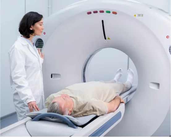
When we talk about treatment for cranial surgery, we’re referring to the set of procedures that give neurosurgeons access to the brain by opening the skull. People often call it brain surgery for short. The reasons for needing such a serious intervention are varied: doctors may remove a tumor, repair a bleeding aneurysm, clear away clotted blood after a traumatic injury, install a shunt for hydrocephalus, or even implant electrodes to help control epilepsy. Regardless of the specific diagnosis, many of these conditions can be life-threatening or lead to permanent disability if left untreated, making surgery a necessary option for many patients.
Over the past couple of decades, the field has made remarkable strides. Surgeons used to rely mostly on their own eye and steady hand; now they have powerful microscopes, three-dimensional brain maps, and even robotic assistants guiding delicate instruments with an accuracy measured in millimeters. These advances mean smaller incisions, less pressure on surrounding tissue, shorter hospital stays, and—perhaps most important—better long-term outcomes. A person who once faced weeks of recovery after a large craniotomy may today walk out of the operating room with little more than a small scar above the ear.
Because cranial surgery involves such complex decisions and cutting-edge technology, it helps to have a clear picture of what the treatment journey looks like from start to finish.
Understanding the treatment for cranial surgery is crucial for patients and their families to navigate the complexities involved in the process.
When Treatment for Cranial Surgery Becomes Necessary
Tumors are arguably the most well-known reason people end up on the neurosurgeon’s schedule. Whether a growth is benign or malignant, surgical resection often remains the best way to relieve pressure, remove active disease, and provide tissue for a definitive diagnosis. By taking out the bulk of a tumor, surgeons can frequently reduce headaches, uncontrolled seizures, weakness, and other symptoms that stem from mass effect on surrounding brain cells.
- Traumatic Brain Injury (TBI) :- When someone suffers a serious head injury—whether from a car crash, a fall, or a sports accident—the brain can bleed, swell, or cause pieces of the skull to fracture. In these tough scenarios, surgeons may open the skull to relieve pressure, clear out blood clots, or patch up broken bone.
- Hydrocephalus :- Hydrocephalus occurs when too much cerebrospinal fluid collects inside the brain, causing dangerous pressure to build up. A reliable fix is to place a shunt that redirects the extra fluid away from the head so the pressure returns to a safer level.
- Aneurysms and Arteriovenous Malformations (AVMs) :- An unruptured aneurysm or AVM can be a ticking time bomb for a brain bleed. To stop that from happening, neurosurgeons might clip the base of an aneurysm, coil its inside with tiny metal spirals, or excise the malformed vessel entirely.
- Epilepsy Surgery :- Many people control their seizures with medication, but those who don’t sometimes find relief through surgery. Doctors will identify the precise spot inside the brain where the seizures start, then carefully remove that section to give the patient a better shot at a seizure-free life.
- Infections or Abscesses :- Brain infections, like abscesses, can be stubborn—they might refuse to budge, even with powerful antibiotics. When that happens, surgeons can make a small opening to drain the pus, clearing the way for the patient to recover. Because every situation is different, surgeons base their choice for cranial surgery on a thorough neurological exam, detailed MRI or CT images, and a candid discussion with a seasoned neurosurgeon.
Types of Cranial Surgery and Treatment Options
Thinking about brain surgery can be nerve-wracking, but knowing the main types can make the process feel a little clearer. Here are the most common procedures you might hear about.
- The craniotomy is probably the surgery most people picture when they think of brain operations. The surgeon lifts a part of the skull to reach the brain directly. This approach lets doctors remove tumors, clip aneurysms, and handle a variety of other issues. Once the work is done, the section of bone is carefully replaced and secured.
- Minimally invasive neurosurgery, sometimes called keyhole surgery, has become a popular alternative for many conditions. By making only small incisions and relying on cameras and special tools, surgeons can get to brain areas that once demanded large openings. Procedures like tumor resections, biopsies, and deep-brain stimulation (DBS) are done this way, allowing for shorter hospital stays and quicker recoveries.
- Endoscopic cranial surgery takes the concept of minimal incision even further. Here, a flexible tube equipped with a camera slides into the body through natural passages, most commonly the nose. This method works well for pituitary tumors, certain cysts, and cerebrospinal fluid (CSF) leaks, since it avoids external scars altogether.
- Stereotactic surgery pairs advanced imaging with a specially designed frame to pinpoint lesions hiding deep within the skull. Whether the goal is targeted radiotherapy or a precise biopsy, the computer-guided system delivers focused energy or needles with remarkable accuracy.
Finally, there are situations when keeping a patient awake is the safest choice. Awake brain surgery allows the surgical team to monitor speech, vision, and movement in real time, helping them steer clear of vital brain areas while still performing critical repairs.
Sophisticated Tools Changing Cranial Surgery
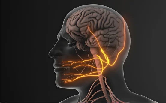
Today’s cranial surgery looks very different from just a few years ago, thanks to a suite of cutting-edge technologies that allow surgeons to work with fresh accuracy. Intraoperative scans, whether MRI or CT, let the surgical team peer inside the skull while the patient is still on the table, showing any last-minute shifts that may have occurred. Neuro-navigation systems, much like GPS for the brain, guide instruments to the exact spot, helping reduce damage to healthy tissue. Robotic arms bring steadiness to delicate tasks that test even the steadiest human hand. Surgeons also rely on detailed 3D brain maps that outline vital speech and movement zones, so they know where to cut and where to hold back. Artificial intelligence tools quietly analyze data, suggesting the best approach based on patterns gleaned from thousands of earlier cases. Altogether, these advances are making even the most challenging neurosurgeries safer than ever.
What to Expect After the Operation
How quickly someone bounces back from cranial surgery hinges on the procedure itself, along with factors such as age, general health, and the reason for the operation. Right after the operation, most patients are taken to the intensive care unit, where staff keep close watch for at least a day. After that, they are moved to a regular ward, and the total hospital stay usually falls somewhere between five days and two weeks, depending on progress. Once home, many will work with physical therapists to regain strength, occupational therapists to tackle daily tasks, and sometimes speech therapists or cognitive specialists if communication or memory is affected. Medically, recovery often includes a regimen of painkillers, anti-seizure drugs, and possibly antibiotics, so follow-up visits with the neurosurgeon to adjust dosages and check incisions are very important. Life outside the hospital may require some calming adjustments—doctors typically recommend steering clear of heavy lifting, while a balanced diet, stress-relieving routines, and light exercise, once cleared, can help the healing process move along nicely.
Risks and Complications of Cranial Surgery
No matter how advanced the tools have become, brain surgery is still delicate work. Patients should be aware of a handful of potential problems that surgeons keep a close eye on. Infection at the incision site or deeper within the skull can sometimes occur, though sterile techniques and antibiotics greatly reduce this risk. Because opening the skull disturbs blood vessels, some bleeding—either outside or inside the brain—remains a possibility. Surgeons manage this with careful technique and, when necessary, additional procedures. Brain swelling, or edema, is another concern; it can press on delicate structures and may necessitate further monitoring in an intensive care unit. Seizures after surgery, though often temporary, are not uncommon and can usually be controlled with medication. Finally, some patients may notice subtle changes in thinking, memory, or movement, which are typically transient but can occasionally be long-lasting.
Choosing an experienced surgical team and a well-equipped hospital can minimize these risks.
Conclusion
Still, the upside of modern cranial work is hard to overlook. Operations that once carried high mortality rates are now routine, and procedures have expanded from traditional craniotomies to minimally invasive endoscopies and even robotic-assisted techniques. Such innovations allow surgeons to tailor each step of the surgery to the patient’s individual anatomy. If you or a loved one faces this crossroads, talking through the case with a board-certified neurosurgeon—and perhaps seeking a second opinion—can make the path forward feel less daunting. Coupled with prompt treatment, cutting-edge imaging, and attentive post-operative care, today’s approach gives many patients the best possible chance for recovery.