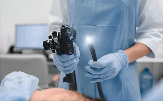
Bronchoscopy is an important procedure that allows physicians to look directly into a patient’s airways and lungs using a slender tube known as a bronchoscope. Over time, the technique has been refined, and there are now several different variations of bronchoscopy surgery, each tailored to address particular health concerns. Understanding the types of bronchoscopy surgery may help patients feel less anxious and better prepared for the experience.
Many doctors turn to bronchoscopy when someone shows symptoms such as a lingering cough, repeated lung infections, or signs of possible lung disease. The procedure not only gives the medical team a clear view of what is happening inside the chest, but it can also deliver treatment on the spot.
Understanding the Types of Bronchoscopy Surgery
Understanding the different types of bronchoscopy surgery can provide insight into the appropriate treatment options available for various lung conditions.
By exploring the various types of bronchoscopy surgery, patients can gain valuable insights into the procedures that may be recommended by their healthcare providers.
So why might a physician order this test in the first place? There are several strong reasons, including:
- Identifying diseases such as tuberculosis, pneumonia, or various forms of lung cancer
- Exploring chronic complaints such as wheezing, ongoing cough, or blood in the sputum
- Extracting a foreign object that has accidentally entered the airway
- Clearing blockages or stopping unexpected bleeding in the bronchial tubes
- Collecting tissue samples or washing fluid from the lungs for lab study
- Keeping tabs on infections or conditions like interstitial lung disease
Because bronchoscopy is relatively minimally invasive, it can often replace more extensive surgical options, making life easier for both patients and doctors.
Types of Bronchoscopy Surgery
Doctors generally rely on two main forms of bronchoscopy: flexible bronchoscopy and rigid bronchoscopy. Each approach has its own set of characteristics, preferred applications, and advantages, tailored to the specific lung issue being treated.
Flexible Bronchoscopy
Flexible bronchoscopy has become the standard option that many clinicians reach for. A thin, pliable tube fitted with a tiny camera and light is slipped through the nose or mouth and carefully threaded down into the airways.
Common Uses of Flexible Bronchoscopy
- Gathering tissue or fluid samples through biopsy or lavage
- Investigating persistent coughs, lung infections, or unusual chest X-ray results
- Identifying tumors, areas of inflammation, or obstructed passages
- Placing stents or delivering laser treatment directly in the airway
Key Advantages
- Typically needs only local numbing or mild sedation
- Can often be done in an outpatient setting with a short recovery
- Provides high-resolution video that aids precise diagnosis
- Reaches smaller airways for a closer look at subtle problems
Rigid Bronchoscopy
- In contrast, rigid bronchoscopy employs a straight, durable metal tube that offers a wider working space. Because of this design, the procedure is carried out under general anesthesia and is usually reserved for therapeutic interventions rather than initial diagnosis.
Rigid bronchoscopy plays a vital role in a number of challenging airway situations. First, it provides a reliable way to extract large foreign bodies that may accidentally block a person’s throat or windpipe, ensuring the airway can be cleared quickly. Second, when a patient is experiencing heavy bleeding in the airway, the rigidity of the instrument gives the doctor the leverage needed to apply interventions that can stop the flow. The same stability makes it useful for removing bulky tumors or placing stents, as well as for opening up severely narrowed or obstructed passages where other tools might struggle.
One of the biggest advantages of rigid bronchoscopy is its superior control during emergencies. Because the scope is solid, surgeons can grasp and maneuver obstructions much more confidently than with flexible instruments. This feature is crucial in trauma cases or in patients whose central airways are compromised by large growths.
In addition to the traditional rigid and flexible approaches, the field has developed several cutting-edge bronchoscopic techniques that cater to specific diagnostic and treatment needs. Electromagnetic navigational bronchoscopy, for instance, uses a combination of CT scans and GPS-like guidance to steer toward small, difficult lung nodules that would be nearly impossible to reach otherwise. This innovation is especially helpful when the goal is to spot early lung cancers. Its minimally invasive nature means patients can avoid the larger incisions traditionally required for such deep biopsies.
Another specialized option is endobronchial ultrasound, or EBUS bronchoscopy. By integrating ultrasound imaging directly into a flexible bronchoscope, physicians can peer around the air passages to view nearby lymph nodes and other structures. While they look, a tiny needle can pass through the scope to take biopsies on the spot, sparing the patient the downtime of an open surgical procedure. Both navigational bronchoscopy and EBUS illustrate how advances in imaging and instrumentation continue to refine airway care.
Uses
Staging lung cancer
- Diagnosing swollen lymph nodes in the chest
- Assessing cases of tuberculosis or sarcoidosis
Auto fluorescence bronchoscopy, or AFB, highlights very early cancerous and precancerous shifts in bronchial tissue. By shining a special blue light onto the airways, the abnormal cells fluoresce and stand out for the doctor. This technique is particularly valuable for spotting lung cancer before it can be seen with standard methods.
Procedure overview
Beforehand, patients are usually asked to avoid food and drinks for six to twelve hours. Flexible bronchoscopy is done with a local anesthetic sprayed into the throat, while a rigid scope sometimes requires general anesthesia. To help with relaxation, the team frequently gives sedatives or medication through an IV line.
Once the procedure begins, a thin bronchoscope slides in through the nose or mouth. The physician not only scans the airways but may also collect tissue samples or carry out small treatments on the spot. Overall, the entire process takes about thirty minutes to one hour.
Afterward, patients are kept in a recovery area for a few hours. They might notice a sore throat, a slight cough, or trace amounts of blood in their saliva, all of which are usually mild and temporary. Most people are back to normal daily activities within a day. Pathology results from biopsies, if taken, typically arrive in a few business days.
As with any procedure, bronchoscopy is generally safe but not risk-free. Possible issues include light bleeding at the biopsy site, infection, a brief episode of wheezing, and, in very rare cases, a pneumothorax or collapsed lung. Always heed your doctor’s advice and contact them right away if you experience severe or unusual symptoms afterward.
Conclusion
Getting a grip on the different types of bronchoscopy is important for anyone facing the procedure, whether you’re the patient or a family member trying to help. From flexible bronchoscopy used mainly for diagnosis to the sturdier rigid version that often tackles more complicated issues, these procedures are vital tools for sorting out lung problems big and small.
Modern technology has really shifted the landscape. Gadgets like navigational bronchoscopy, endobronchial ultrasound, and automated flexible bronchoscopy have made the process safer, more precise, and much less taxing on the body. If your doctor has suggested you have one done, take a moment to talk it over with an experienced pulmonologist so you know exactly why a particular type is recommended for you.