Craniotomy is a surgical procedure where a portion of the skull is temporarily removed to access the brain. It is a common yet highly specialized technique used to treat various neurological conditions such as brain tumors, aneurysms, traumatic brain injury, and epilepsy. But not all craniotomies are the same. There are several types, each designed for a specific purpose based on the location of the problem and the complexity of the case.
Understanding the different types of craniotomy surgeries can help patients and caregivers better prepare for treatment, discuss options with their neurosurgeon, and feel more informed about the process and recovery.
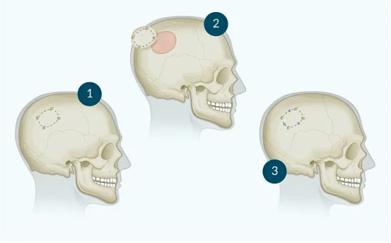
Types of Craniotomy Surgery
- Traditional Craniotomy :- The traditional or standard craniotomy is the most widely performed technique. In this procedure, a section of the skull, known as a bone flap, is removed to allow the surgeon to access the brain. Once the surgery is complete, the bone is replaced and secured with plates or screws. This type of craniotomy is typically done under general anesthesia and is used for a variety of conditions including tumors, blood clots, and brain swelling.
- Awake Craniotomy :- Awake craniotomy is a specialized procedure often used when the tumor or abnormality is located near brain regions that control important functions like speech, movement, or memory. During this surgery, the patient is kept awake but pain-free. They interact with the surgical team, helping the neurosurgeon monitor brain function in real time. This approach minimizes the risk of damaging essential brain areas while allowing for more precise tumor removal or seizure focus resection.
- Keyhole Craniotomy (Minimally Invasive Craniotomy) :- Keyhole craniotomy involves making a much smaller opening in the skull compared to a traditional craniotomy. The surgeon uses advanced imaging and tools to reach the targeted area with minimal disruption to surrounding brain tissue. This technique is often used for smaller tumors, pituitary lesions, or certain aneurysms. Because the incision is smaller, patients generally experience shorter hospital stays, less pain, and faster recovery.
- Endoscopic Craniotomy :- In an endoscopic craniotomy, a tiny camera (endoscope) is inserted through a small skull opening to guide the surgeon. This type of craniotomy is particularly useful for deep-seated brain tumors or lesions. It allows for high-definition visualization and improved access with minimal impact on surrounding tissues. It is a preferred method when the goal is to reduce trauma while maintaining surgical precision.
- Supraorbital Craniotomy (Eyebrow Craniotomy) :- This minimally invasive technique involves making an incision above the eyebrow to access tumors or abnormalities in the front part of the brain. It provides access through the natural skull base and avoids large incisions across the scalp. Supraorbital craniotomy is often used for tumors in the frontal lobes or around the optic nerve and can result in reduced scarring and a quicker return to daily activities.
- Orbitozygomatic Craniotomy :- This more extensive type of craniotomy involves removing not just a portion of the skull, but also parts of the bones around the eye socket and cheekbone. It provides greater access to deep and complex regions of the brain, particularly in cases involving large or deeply located tumors at the base of the skull. Due to its complexity, this procedure is typically reserved for high-risk or hard-to-reach brain lesions.
- Pterional Craniotomy (Frontotemporal Craniotomy) :- A pterional craniotomy, also known as a frontotemporal approach, involves accessing the brain through the temple area. This technique is frequently used for treating aneurysms, tumors near the optic nerves, and lesions in the frontal and temporal lobes. The pterional approach allows surgeons to work around critical brain structures while maintaining a wide field of view.
- Extended Bifrontal Craniotomy :- This is a more aggressive and larger craniotomy performed for large tumors or multiple abnormalities located in both frontal lobes. It involves removing bone from both sides of the forehead to gain broad access to the front part of the brain. It’s often used for complex tumors that are otherwise difficult to reach with standard approaches.
Conclusion
Craniotomy is not a one-size-fits-all procedure. Neurosurgeons choose the most appropriate type based on the condition being treated, the location of the lesion, and the overall goal of the surgery. Whether it’s a minimally invasive keyhole approach or a complex bifrontal technique, every type of craniotomy is designed to offer the safest, most effective path to recovery.
Open communication with your neurosurgical team, along with a clear understanding of the planned approach, can make the experience less intimidating and more manageable. With advancements in technology and surgical methods, craniotomy today is safer and more precise than ever before.