Introduction
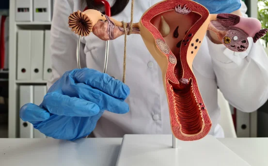
Dacryocystorhinostomy (DCR) is a surgical procedure performed to treat blockage of the nasolacrimal duct, which is responsible for draining tears from the eye into the nose. When this duct becomes obstructed, patients experience excessive tearing (epiphora), recurrent eye infections, swelling near the inner corner of the eye, and discomfort. DCR creates a new drainage pathway between the lacrimal sac and the nasal cavity, bypassing the blocked duct and restoring normal tear flow.
Over the years, advancements in surgical techniques have led to different types of dacryocystorhinostomy, each suited to specific patient needs and anatomical conditions. Understanding these types helps patients and caregivers make informed decisions regarding treatment.
What Is Dacryocystorhinostomy?
Dacryocystorhinostomy is commonly recommended when conservative treatments such as medications or lacrimal probing fail to resolve tear duct obstruction. The procedure aims to permanently relieve symptoms by allowing tears to drain freely into the nasal cavity. DCR can be performed using external or internal approaches, with or without advanced instruments such as endoscopes or lasers. The choice of technique depends on factors like age, severity of blockage, nasal anatomy, history of previous surgeries, and surgeon expertise.
Types of Dacryocystorhinostomy
- External Dacryocystorhinostomy :- External DCR is the traditional and most widely practiced form of dacryocystorhinostomy. It involves making a small incision on the side of the nose near the inner corner of the eye to directly access the lacrimal sac.
Key Features
- A skin incision allows direct visualization of the lacrimal sac
- A bony opening is created to connect the lacrimal sac to the nasal cavity
- Often performed under local or general anesthesia
Advantages :- External DCR has a high success rate, typically above 90%. Surgeons have direct access to the anatomy, allowing precise removal of the obstruction. It is especially effective in complex cases or revision surgeries.
Limitations :- The main drawback is the presence of a small facial scar, although it usually fades over time. Recovery may be slightly longer compared to minimally invasive methods.
- Endoscopic Dacryocystorhinostomy :- Endoscopic DCR is a minimally invasive technique performed through the nasal cavity using an endoscope. No external incision is required, making it cosmetically appealing.
Key Features
- Performed entirely through the nose
- Uses a nasal endoscope for visualization
- Commonly done under general anesthesia
Advantages :- Endoscopic DCR avoids facial scarring and preserves normal anatomy. Patients typically experience less pain, minimal bleeding, and faster recovery. It is also beneficial for patients with associated nasal conditions such as deviated nasal septum or nasal polyps.
Limitations :- The success rate may be slightly lower than external DCR in some cases. It requires specialized equipment and an experienced surgeon for optimal outcomes.
- Laser-Assisted Dacryocystorhinostomy :- Laser-assisted DCR uses laser energy to create the opening between the lacrimal sac and the nasal cavity. This technique can be performed via an endonasal or transcanalicular approach.
Key Features
- Uses diode or Nd:YAG lasers
- Minimally invasive with reduced tissue trauma
- Often performed as a day-care procedure
Advantages :- Laser DCR offers minimal bleeding, shorter operating time, and quicker recovery. It is particularly suitable for patients seeking a less invasive option and faster return to daily activities.
Limitations :- Laser DCR may have a slightly lower long-term success rate compared to conventional methods. It may not be ideal for patients with severe or complex obstructions.
- Transcanalicular Dacryocystorhinostomy :- Transcanalicular DCR is performed by inserting fine instruments through the natural tear drainage canal (canaliculus) of the eye.
Key Features
- No skin incision or nasal opening
- Access through the tear duct itself
- Can be laser-assisted or mechanical
Advantages :- This technique is cosmetically excellent, as there are no visible scars. It is less invasive and associated with minimal postoperative discomfort.
Limitations :- The procedure has limited visibility and may not be suitable for all types of obstructions. Success rates depend heavily on patient selection and surgeon experience.
- Revision Dacryocystorhinostomy :- Revision DCR is performed when a previous dacryocystorhinostomy has failed due to scarring, closure of the new drainage pathway, or infection.
Key Features
- Can be external or endoscopic
- Focuses on reopening or enlarging the blocked passage
- Often requires stent placement
Advantages :- Revision DCR provides a second opportunity to restore tear drainage and relieve persistent symptoms. With proper evaluation, success rates remain high.
Limitations :- Revision surgeries can be more technically challenging due to scar tissue and altered anatomy.
- Pediatric Dacryocystorhinostomy :- In children, tear duct obstruction is often congenital. Pediatric DCR is considered only when conservative treatments and probing fail.
Key Features
- Performed under general anesthesia
- Tailored to delicate pediatric anatomy
- Usually endoscopic or external
Advantages :- Pediatric DCR effectively resolves chronic tearing and recurrent infections, improving comfort and eye health in children.
Limitations :- Surgery is typically delayed until the child reaches an appropriate age, unless complications arise earlier.
Choosing the Right Type of Dacryocystorhinostomy
The choice of dacryocystorhinostomy depends on several factors, including the cause and location of the blockage, patient age, cosmetic concerns, previous surgeries, and available surgical expertise. A thorough evaluation by an ophthalmologist or ENT specialist is essential to determine the most suitable approach.
Conclusion
Dacryocystorhinostomy is a highly effective solution for chronic tear duct obstruction. With multiple types of DCR available ranging from traditional external techniques to modern endoscopic and laser-assisted procedures patients now have safer, less invasive, and cosmetically favorable options. Early diagnosis and timely surgical intervention can significantly improve quality of life by relieving tearing, preventing infections, and restoring normal tear drainage.