Scleral buckling surgery has been performed for decades and continues to be an effective solution for certain types of retinal tears and detachments, especially rhegmatogenous retinal detachment, the most common form caused by a break or hole in the retina.
Retinal detachment is a serious eye condition that can lead to permanent vision loss if not treated promptly. One of the most effective and time-tested surgical treatments for this condition is types of scleral buckling surgery. Understanding the different types of scleral buckling procedures can help patients make informed decisions and reduce anxiety surrounding surgery.
What Is Scleral Buckling Surgery?
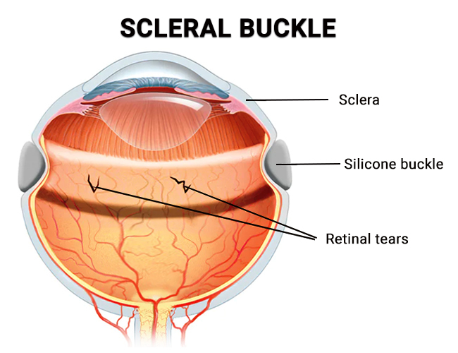
Scleral buckling is a surgical procedure used to repair retinal detachment, which occurs when the retina—the light-sensitive tissue at the back of the eye pulls away from the underlying support tissue.
The surgery involves placing a small silicone or sponge-like material (the “buckle”) on the sclera (the white outer layer of the eye). This buckle gently presses the wall of the eye inward to relieve the pulling on the retina, allowing it to reattach and heal.
When Is Scleral Buckling Recommended?
Scleral buckling is primarily used for rhegmatogenous retinal detachment (RRD), which is caused by a tear or hole in the retina. The procedure may be recommended if :-
-
The detachment is located in the peripheral retina
-
The retinal tear is relatively small and identified early
-
The patient is younger or highly myopic
-
There is no significant scar tissue (proliferative vitreoretinopathy)
Different Types of Scleral Buckling Surgery
There are several types of scleral buckling procedures. The choice depends on the size, location, and severity of the retinal tear or detachment, as well as the surgeon’s preference and patient-specific factors.
Here are the major types of scleral buckling surgeries :-
Segmental Buckle Surgery
Segmental buckling is used when there’s a localized retinal tear or detachment. In this procedure, a silicone sponge or rubber element is sewn onto a small section of the sclera directly underneath the retinal tear.
Key Features :-
-
Treats localized retinal tears
-
Less invasive than a full buckle
-
Typically performed under local anesthesia
Advantages :-
-
Less disruption to the shape of the eye
-
Reduced risk of refractive changes post-surgery
Encircling Buckle Surgery
In encircling scleral buckle surgery, a band is placed around the entire circumference of the eye (like a belt). This band uniformly indents the eyeball to relieve traction from multiple retinal tears or widespread retinal degeneration.
Key Features :-
-
Used for multiple or extensive tears
-
Common in complex or recurrent retinal detachments
Advantages :-
-
Supports the entire retina
-
Effective for more severe or large-area detachments
Considerations :-
-
May induce myopia due to change in eye shape
-
Usually performed under general anesthesia
Radial Buckle Surgery
Radial buckling involves placing a buckle perpendicular to the retina, in a radial direction, to support specific types of tears like horseshoe-shaped tears.
Key Features :-
-
Ideal for retinal breaks at the posterior pole
-
Supports anterior-posterior traction
Advantages :-
- Precise support for specific tear orientations
- Useful when tears are not located near the ora serrata
Circumferential Buckle Surgery
Unlike the encircling buckle that goes all the way around, a circumferential buckle is a partial band placed along a specific meridian of the eye.
Key Features :-
-
Used when multiple breaks occur in the same quadrant
-
Customizable to the location of retinal tears
Advantages :-
-
Less risk of changing eye shape compared to a full band
-
Reduced surgical time and recovery
Combined Scleral Buckling with Vitrectomy
In some cases, especially when there’s vitreous hemorrhage or traction, scleral buckling is combined with pars plana vitrectomy (PPV).
Key Features :-
-
Used in complicated or recurrent detachments
-
Removes vitreous gel while supporting the retina with a buckle
Advantages :-
-
Provides a multi-faceted repair approach
-
Improves outcomes in complex retinal cases
How to Choose the Right Type of Scleral Buckling Surgery?
The decision on which type of scleral buckle to use depends on several clinical factors :-
-
Location of the tear :- Peripheral vs posterior
-
Number of tears :- Single or multiple
-
Severity of detachment
-
Patient age and eye anatomy
-
Previous eye surgeries or trauma
-
Presence of other eye conditions
An experienced retinal surgeon will assess your condition through dilated eye exams, ultrasound imaging, and OCT scans before recommending the most suitable technique.
What to Expect During and After the Surgery?
Preoperative Steps :-
-
Comprehensive eye examination
-
Retinal imaging to locate tears
-
Counseling about anesthesia and procedure
During Surgery :-
-
Usually takes 1–2 hours
-
Local or general anesthesia depending on case
-
Cryopexy or laser photocoagulation may be used to seal retinal breaks
Postoperative Recovery :-
-
Eye patch and medications to reduce inflammation and infection
-
Blurred vision is normal for a few weeks
-
Avoid strenuous activity or heavy lifting
-
Follow-up visits to monitor reattachment
Risks and Complications
Though scleral buckling is a safe and well-established procedure, like any surgery, it carries some risks :-
-
Infection or bleeding
-
Double vision (temporary)
-
Re-detachment of the retina
-
Increased intraocular pressure
-
Induced myopia (especially with encircling buckle)
Early diagnosis and choosing the right type of surgery significantly reduce the likelihood of complications.
Advantages of Scleral Buckling
-
High success rates for rhegmatogenous retinal detachments
-
Long-lasting repair with minimal recurrence
-
Preserves natural vitreous in younger patients
-
Can be customized to specific retinal break locations
Conclusion
Scleral buckling surgery remains a gold standard for treating certain types of retinal detachment, especially in younger patients or those with peripheral retinal breaks. The various types segmental, encircling, radial, circumferential, and combined with vitrectomy allow for a tailored approach to each patient’s unique condition.