What Is Vitrectomy Surgery?
Vitrectomy eye surgery is a simple procedure that takes out the jelly-like vitreous humor from the center of the eye. Surgeons often use it when the retina is torn, or when cloudy bleed, scar tissue, or tiny pieces of debris block clear sight. Doctors choose vitrectomy when the eye’s gel or the retina itself causes big problems. Here are the main reasons they go in:
Understanding the different types of vitrectomy surgery is crucial for patients considering this procedure.
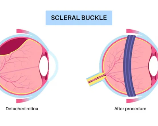
Retinal Detachment
When the retina peels away from its wall, time runs out. A vitrectomy lets the surgeon push it back with a quick laser, then seats a tiny gas bubble to hold everything in place.
Macular Hole or Macular Pucker
- Small tears or tight wrinkles form on the spot that makes detail clear. Removing the gel gives the doctor room to patch them and often sharpens straight-ahead vision.
Diabetic Retinopathy
- High blood sugar can ruin eye vessels, leaking blood into the jelly. The clean-up stops the flood and keeps diabetes from darkening the field of sight.
Vitreous Hemorrhage
- Fresh bleeding inside the gel turns clear space red, blocking useable view. A quick vitrectomy sweeps the blood away and lets light travel again.
Eye Floaters
- If floaters keep bothering you, the doctor might suggest a partial or full vitrectomy to clear them out.
Types of Vitrectomy Surgery
Surgeons pick one of three main approaches based on where the problem is and how serious it is. The choice depends on the patients exact symptoms and the condition of the eye.
Anterior Vitrectomy
Overview:
This quick fix is done when vitreous gel slips into the front chamber of the eye, often because of a tear during cataract surgery.
Indications:
- Posterior capsule tear
- Lens fragments out of place
- Vitreous pushing forward
Procedure:
The surgeon makes a tiny cut at the front of the eyeball. A small probe slides in, scoops out the loose gel, and brings back clearer sight.
Benefits:
- Keeps trouble from spreading
- Stabilizes the front chamber
- Lowers odds of infection or swelling
Posterior Pars Plana Vitrectomy (PPV)
Overview:
Pars plana vitrectomy is the go-to version. The surgeon drills mini ports in the pars plana, the flat section in the whites of the eye, so he can reach the jelly cavity and the retina.
Indications:
- retinal detachment
- macular hole
- diabetic retinopathy
- vitreous hemorrhage
Procedure:
The procedure is quick and uses tiny, specialized tools while the患者 is under either local or light general anesthesia. The cloudy vitreous gel is gently suctioned out, and a laser or freezing probe seals the retina. A bubble of gas or a small plug of silicone oil is placed so the tissue stays smooth while new layers grow.
Benefits:
- restores retinal health
- improves vision outcomes
- enables treatment of complex retinal conditions
Endoscopic Vitrectomy
Overview:
endoscopic vitrectomy uses a small bendable camera when the inside of an eye is too cloudy for standard lights-its a lifesaver after serious injury or infection.
Indications:
- severe eye trauma
- endophthalmitis (eye infection)
- dense corneal opacity or media opacities
Procedure:
a miniature scope and hand-held instruments slide through a tiny opening, sending live video to the surgeons screen so every cut can be aimed exactly. this technique stays reserved for the most delicate cases.
Benefits:
- Lets doctors treat problems in hard-to-reach parts of the eye
- Can keep patients out of bigger, scarier surgery rooms
- Works well for tricky trauma injuries
Technological Advances in Vitrectomy
Today surgeons cut the eye with tiny 25-gauge or 27-gauge tools, use sharper cameras, and feed tissue out with safer cutters. Because of this patients enjoy:
- Faster recovery time
- Reduced post-op complications
- Higher success rates
- Wide-angle lenses and fluorescein dye now give the surgeon a brighter, wider map to follow.
Recovery After Vitrectomy Surgery
How long eyes heal depends on the problem and which vitrectomy was done. Until vision settles, patients should follow these simple rules:
- Positioning: Face-down sitting is key if the team leaves a gas bubble.
- Eye Drops: Expect stinging from anti-inflammatory and antibiotic drops.
- Follow-Up: Multiple quick clinic stops check the eye step by step.
- Restrictions: Stay out of planes or high altitude as long as the bubble floats.
Conclusion
Vitrectomy is one of ophthalmologys go-to moves, giving light and life back to patients with serious eye disease. With options like anterior vitrectomy, pars plana vitrectomy, or endoscopic gear, the surgeon picks the best fit for each case.
If you notice quick changes like blurred vision, new floaters, or anything strange with your retina, see a good eye doctor ASAP. Catching stuff early makes a huge difference in how well you see later.
From torn retinas and diabetes damage to macular problems, vitrectomy remains a game-changer for thousands, fixing vision and boosting everyday life.