Hydrocephalus is a serious medical condition characterized by an abnormal accumulation of cerebrospinal fluid (CSF) within the brain’s ventricles (fluid-filled cavities). This excessive buildup leads to increased intracranial pressure, which can damage brain tissues and cause a variety of neurological symptoms. If left untreated, hydrocephalus can lead to permanent brain damage or even become life-threatening.
Hydrocephalus surgery is a crucial intervention designed to relieve this pressure and manage the condition effectively. In most cases, surgical treatment involves diverting or draining the excess CSF to another part of the body where it can be absorbed. The procedure helps restore the balance of fluid in the brain and prevents further complications.
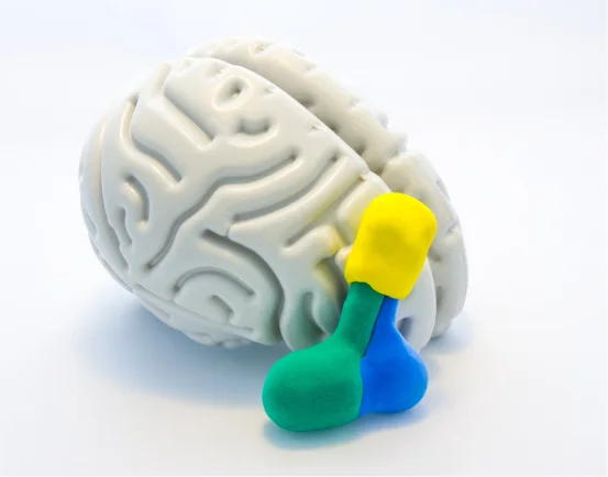
Understanding What is Hydrocephalus Surgery?
Cerebrospinal fluid is a clear, protective fluid that surrounds the brain and spinal cord. It plays a critical role in cushioning the brain, removing waste products, and delivering nutrients. Normally, the body produces and absorbs CSF in a balanced cycle. In hydrocephalus, this balance is disrupted either by overproduction, blockage of flow, or impaired absorption of CSF.
The condition can be congenital (present at birth) or acquired later due to injury, infection, tumor, or hemorrhage. It can affect people of all ages but is especially common in infants and older adults.
Common Causes of Hydrocephalus
Hydrocephalus can develop for various reasons, including:
- Congenital malformations such as aqueductal stenosis or Dandy-Walker syndrome
- Brain hemorrhage, especially in premature infants
- Infections such as meningitis or encephalitis
- Head trauma
- Brain tumors blocking CSF flow
- Normal Pressure Hydrocephalus (NPH) in older adults
In all cases, surgical treatment is the primary option to manage the condition.
Why is Hydrocephalus Surgery Needed?
The brain is enclosed within the rigid skull, so any increase in fluid volume causes pressure to rise. This intracranial pressure (ICP) can compress delicate brain tissues and lead to serious complications such as:
- Vision problems
- Headaches
- Vomiting
- Seizures
- Developmental delays
- Memory loss or confusion
- Difficulty walking
- In severe cases, coma or death
Surgery is necessary to prevent or reverse damage caused by increased pressure. Medical therapy alone is usually ineffective.
Types of Hydrocephalus Surgery
There are two primary surgical approaches for treating hydrocephalus:
Ventriculoperitoneal (VP) Shunt Surgery
This is the most commonly performed surgery for hydrocephalus. A VP shunt is a flexible, silicone tube system implanted under the skin to divert excess CSF from the brain to another part of the body—usually the peritoneal cavity in the abdomen, where the fluid is absorbed naturally.
How it Works:
- A small hole is made in the skull to access a brain ventricle.
- A catheter (tube) is inserted into the ventricle to collect the CSF.
- The tube is connected to a valve that regulates fluid flow.
- The other end of the tube is tunneled under the skin and inserted into the abdomen.
The shunt system continuously drains CSF as needed, reducing intracranial pressure and maintaining balance.
Advantages:
- Long-standing and widely used procedure
- Effective in infants, children, and adults
- Can be adjusted or replaced as needed
Limitations:
- Shunts can malfunction or become infected
- Multiple surgeries may be required over a lifetime
- Regular follow-up is essential
Endoscopic Third Ventriculostomy (ETV)
ETV is a less invasive alternative to shunt placement, typically used for non-communicating (obstructive) hydrocephalus, where the flow of CSF is blocked within the ventricles.
How it Works:
- A neurosurgeon uses an endoscope (thin camera) to enter the brain’s third ventricle.
- A small hole is made at the bottom of the ventricle.
- This hole allows CSF to bypass the blockage and flow into the natural absorption pathways.
Advantages:
- No foreign device (shunt) is needed
- Lower long-term complication rates
- Preferred in cases of aqueductal stenosis
Limitations:
- Not suitable for all types of hydrocephalus
- Requires high surgical precision
- Risk of bleeding or damage to brain structures
In some cases, ETV may be combined with choroid plexus cauterization (CPC), a technique used to reduce CSF production, especially in infants.
What to Expect Before Surgery
Before hydrocephalus surgery, the patient undergoes thorough evaluation including:
- Neurological examination
- Brain imaging (MRI or CT scan)
- Pressure monitoring (in certain cases)
- Blood tests and infection screening
In infants and children, developmental assessments are also performed. Informed consent is obtained, and the family is educated about the surgical plan, risks, and benefits.
Postoperative Recovery
Recovery from hydrocephalus surgery depends on the patient’s age, the severity of the condition, and the type of surgery performed.
Immediate Post-Surgery Period:
- Monitoring in a hospital setting for 24–72 hours
- Pain management and wound care
- Observation for signs of complications such as infection or shunt malfunction
- Gradual resumption of normal feeding or mobility in infants
Recovery Timeline
- Most patients are discharged within a few days.
- Return to school or work may take 1 to 2 weeks.
- Follow-up imaging is often done within the first month.
- Children may require physical, occupational, or speech therapy depending on pre-surgical symptoms.
Possible Complications
As with any surgery, hydrocephalus surgery carries risks, including:
- Shunt infection: Requires antibiotics and possible replacement
- Shunt malfunction or blockage: Can cause symptoms to return; may need surgical revision
- Over-drainage: Can lead to headaches, subdural hematoma, or slit ventricle syndrome
- Bleeding or damage to brain tissue
- Seizures
- Scar formation around the shunt system
Prompt medical attention is necessary if symptoms like severe headache, vomiting, lethargy, or fever appear after surgery.
Living with Hydrocephalus After Surgery
Many individuals with hydrocephalus go on to lead full and productive lives after surgery. However, long-term follow-up is essential.
- Regular checkups :- To monitor the function of the shunt or ETV.
- Neurodevelopmental support :- For children with delays in speech, learning, or coordination.
- Education and social support :- Schools and caregivers should be aware of the child’s condition.
- Emergency awareness :- Caregivers should know the signs of shunt failure or infection.
The psychological impact of living with a chronic condition can be significant, especially in children and adolescents. Access to counseling or peer support groups may be beneficial.
Conclusion
Hydrocephalus is a complex condition, but it is treatable with timely surgical intervention. Whether through shunt placement or endoscopic techniques like ETV, hydrocephalus surgery plays a vital role in preserving brain function and improving quality of life.
While the journey doesn’t end with surgery, the right combination of medical care, follow-up, and support can help patients manage the condition effectively. Advances in surgical techniques and materials continue to improve outcomes, offering hope to families affected by hydrocephalus around the world.
If you or your loved one is facing a diagnosis of hydrocephalus, consult with a neurosurgeon or pediatric neurologist to explore your treatment options and ensure the best possible care.