Introduction
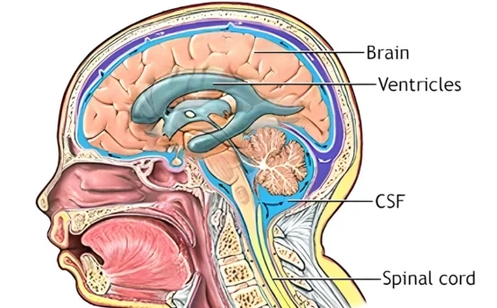
Ventriculoperitoneal (VP) shunt surgery is a critical neurosurgical procedure designed to treat hydrocephalus, a condition characterized by the excessive accumulation of cerebrospinal fluid (CSF) in the brain. This accumulation can lead to increased intracranial pressure, causing severe headaches, vision problems, cognitive difficulties, and even life-threatening complications if left untreated. In this blog, we explore what is ventriculoperitoneal shunt surgery is, how it works, its benefits, risks, and what patients can expect during recovery.
Understanding Hydrocephalus: Why a VP Shunt is Needed
Hydrocephalus occurs when there is an imbalance between the production and absorption of cerebrospinal fluid (CSF) in the brain. CSF surrounds and cushions the brain and spinal cord, but an excess can lead to increased pressure inside the skull.
Common causes of hydrocephalus include
- Congenital conditions, such as spina bifida
- Brain tumors or cysts
- Traumatic brain injury
- Infections like meningitis
- Complications from premature birth
Left untreated, hydrocephalus can cause permanent brain damage. This is where ventriculoperitoneal shunt surgery comes into play.
What is Ventriculoperitoneal Shunt Surgery?
A ventriculoperitoneal (VP) shunt is a medical device that helps drain excess CSF from the brain’s ventricles (fluid-filled cavities) to the peritoneal cavity (the area surrounding the abdominal organs). By diverting the fluid, the shunt relieves intracranial pressure, reducing symptoms and preventing further brain damage.
The procedure is one of the most common treatments for hydrocephalus and can be performed on both children and adults.
Components of a VP Shunt
A VP shunt consists of three main parts
- Ventricular Catheter :- A thin tube inserted into one of the brain’s ventricles to drain excess CSF.
- Valve :- Controls the flow of CSF, ensuring it drains at the correct rate to prevent overdrainage.
- Peritoneal Catheter :- A tube that runs from the valve to the peritoneal cavity, where the CSF is absorbed into the bloodstream.
Modern VP shunts are often programmable, allowing neurosurgeons to adjust the valve pressure without additional surgery.
How VP Shunt Surgery is Performed
VP shunt surgery is typically performed under general anesthesia. The procedure generally involves the following steps:
- Preparation :- The patient is positioned, and the surgical areas on the head and abdomen are sterilized.
- Incision :- Small incisions are made in the scalp and abdomen.
- Insertion of Ventricular Catheter :- The surgeon inserts a catheter into the brain’s ventricle to drain excess CSF.
- Valve Placement :- The valve is connected to the ventricular catheter and placed under the scalp.
- Peritoneal Catheter Placement :- The distal catheter is tunneled under the skin to the abdominal cavity.
- Closure :- All incisions are closed, and the system is tested to ensure proper CSF flow.
The surgery usually takes 1 to 2 hours, depending on the patient’s condition and complexity.
Benefits of Ventriculoperitoneal Shunt Surgery
VP shunt surgery offers multiple benefits for patients suffering from hydrocephalus
- Relieves Intracranial Pressure :- Reduces headaches, nausea, and vomiting caused by excess CSF.
- Improves Cognitive Function :- Can help restore memory, concentration, and overall brain function.
- Prevents Brain Damage :- Protects against long-term complications such as motor impairment and vision loss.
- Improves Quality of Life :- Allows patients to resume normal activities with fewer symptoms.
Risks and Complications
While VP shunt surgery is generally safe, it carries potential risks
- Infection :- Infections in the shunt or surrounding tissue may require removal or replacement.
- Shunt Blockage :- The shunt may become blocked, leading to recurrent symptoms.
- Overdrainage or Underdrainage :- Incorrect CSF flow can cause headaches, subdural hematomas, or continued hydrocephalus symptoms.
- Mechanical Failure :- Rarely, parts of the shunt may malfunction, requiring revision surgery.
Regular follow-ups and imaging studies help detect complications early and ensure the shunt functions properly.
Recovery After VP Shunt Surgery
Post-surgery recovery varies by age and health condition but generally includes
- Hospital Stay :- Most patients stay 3-7 days post-surgery for monitoring.
- Activity Restrictions :- Avoid strenuous activities for a few weeks to allow healing.
- Follow-Up Care :- Regular check-ups and imaging to monitor shunt function.
- Symptom Monitoring :- Watch for headaches, nausea, fever, or swelling around the shunt site.
Children may need long-term monitoring, as they might outgrow the shunt, requiring adjustments or replacements.
Advancements in VP Shunt Technology
Medical advancements have improved VP shunt safety and effectiveness. Some notable developments include
- Programmable Shunts :- Allow valve pressure adjustments non-invasively.
- Antimicrobial Shunts :- Reduce the risk of infection.
- Endoscopic Assistance :- Minimally invasive approaches to improve catheter placement.
These innovations have significantly improved patient outcomes and reduced complications.
When to Consult a Neurosurgeon
Early intervention is critical for managing hydrocephalus. Individuals experiencing persistent headaches, nausea, vision changes, or cognitive issues should consult a neurosurgeon. Pediatric patients showing delayed milestones, enlarged head size, or vomiting may also require evaluation for a VP shunt.
Conclusion
Ventriculoperitoneal shunt surgery is a life-saving procedure for patients with hydrocephalus. By effectively draining excess cerebrospinal fluid, it alleviates symptoms, protects brain function, and significantly enhances quality of life. While risks exist, advances in shunt technology and proper follow-up care make VP shunt surgery a reliable solution for managing this complex neurological condition.